Research Lab Results
-
John Ulatowski Lab
Research in the John Ulatowski Lab explores the regulatory mechanisms of oxygen delivery to the brain and cerebral blood flow. Our work includes developing and applying new techniques and therapies for stroke as well as non-invasive techniques for monitoring brain function, fluid management and sedation in brain injury patients. We also examine the use of novel oxygen carriers in blood. We’ve recently begun exploring new methods for perioperative and periprocedural care that would help to optimize patient safety in the future. -
Gregg Semenza Lab
The Gregg Semenza Lab studies the molecular mechanisms of oxygen homeostasis. We have cloned and characterized hypoxia-inducible factor 1 (HIF-1), a basic helix-loop-helix transcription factor. Current research investigates the role of HIF-1 in the pathophysiology of cancer, cerebral and myocardial ischemia, and chronic lung disease, which are the most common causes of mortality in the U.S.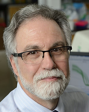
-
Espenshade Lab
The Espenshade Lab uses a multi-organismal and multidisciplinary approach to understand how eukaryotic cells measure insoluble lipids and dissolved gases. We have chosen cholesterol and oxygen as our model molecules, based on their essential roles in cell function and the importance of their proper homeostasis for human health. -
Roy Brower Lab
The Roy Brower Lab conducts clinical trials related to the management of acute respiratory distress syndrome (ARDS). Our research also involves oxygen toxicity, a potentially fatal condition caused by too much supplemental oxygen.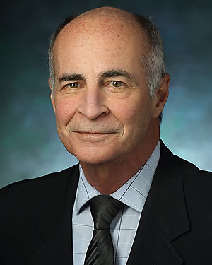
-
Raymond Koehler Lab
Research in the Raymond Koehler Lab explores cerebrovascular physiology and cerebral ischemic injury caused by stroke and cardiac arrest, using protein analysis, immunohistochemistry and histology. We also study ischemic preconditioning, neonatal hypoxic-ischemic encephalopathy and the mechanisms of abnormal cerebrovascular reactivity after ischemia. We 're examining ways to improve tissue oxygenation and seek to better understand the mechanisms that connect an increase in cerebral blood flow to neuronal activity. -
Jun Hua Lab
Dr. Hua's research has centered on the development of novel MRI technologies for in vivo functional and physiological imaging in the brain, and the application of such methods for studies in healthy and diseased brains. These include the development of human and animal MRI methods to measure functional brain activities, cerebral perfusion and oxygen metabolism at high (3 Tesla) and ultra-high (7 Tesla and above) magnetic fields. He is particularly interested in novel MRI approaches to image small blood and lymphatic vessels in the brain. Collaborating with clinical investigators, these techniques have been applied 1) to detect functional, vascular and metabolic abnormalities in the brain in neurodegenerative diseases such as Huntingdon's disease (HD), Parkinson's disease (PD), Alzheimer's disease (AD) and mental disorders such as schizophrenia; and 2) to map brain functions and cerebrovascular reactivity for presurgical planning in patients with vascular malformations, brain tumors and epilepsy. -
James Pekar Lab
How do we see, hear, and think? More specifically, how can we study living people to understand how the brain sees, hears, and thinks? Recently, magnetic resonance imaging (MRI), a powerful anatomical imaging technique widely used for clinical diagnosis, was further developed into a tool for probing brain function. By sensitizing magnetic resonance images to the changes in blood oxygenation that occur when regions of the brain are highly active, we can make ""movies"" that reveal the brain at work. Dr. Pekar works on the development and application of this MRI technology. Dr. Pekar is a biophysicist who uses a variety of magnetic resonance techniques to study brain physiology and function. Dr. Pekar serves as Manager of the F.M. Kirby Research Center for Functional Brain Imaging, a research resource where imaging scientists and neuroscientists collaborate to study brain function using unique state-of-the-art techniques in a safe comfortable environment, to further develop such techniques, and to provide training and education. Dr. Pekar works with center staff to serve the center's users and to keep the center on the leading edge of technology. -
Zaver M. Bhujwalla Lab – Cancer Imaging Research
Dr. Bhujwalla’s lab promotes preclinical and clinical multimodal imaging applications to understand and effectively treat cancer. The lab’s work is dedicated to the applications of molecular imaging to understand cancer and the tumor environment. Significant research contributions include 1) developing ‘theranostic agents’ for image-guided targeting of cancer, including effective delivery of siRNA in combination with a prodrug enzyme 2) understanding the role of inflammation and cyclooxygenase-2 (COX-2) in cancer using molecular and functional imaging 3) developing noninvasive imaging techniques to detect COX-2 expressing in tumors 4) understanding the role of hypoxia and choline pathways to reduce the stem-like breast cancer cell burden in tumors 5) using molecular and functional imaging to understand the role of the tumor microenvironment including the extracellular matrix, hypoxia, vascularization, and choline phospholipid metabolism in prostate and breast cancer invasion and metastasis, with the ultimate goal of preventing cancer metastasis and 6) molecular and functional imaging characterization of cancer-induced cachexia to understand the cachexia-cascade and identify novel targets in the treatment of this condition. -
Machine Biointerface Lab
Dr. Fridman's research group invents and develops bioelectronics for Neuroengineering and Medical Instrumentation applications. We develop innovative medical technology and we also conduct the necessary biological studies to understand how the technology could be effective and safe for people. Our lab is currently focused on developing the ""Safe Direct Current Stimulation"" technology, or SDCS. Unlike the currently available commercial neural prosthetic devices, such as cochlear implants, pacemakers, or Parkinson's deep brain stimulators that can only excite neurons, SDCS can excite, inhibit, and even sensitize them to input. This new technology opens a door to a wide range of applications that we are currently exploring along with device development: e.g. peripheral nerve stimulation for suppressing neuropathic pain, vestibular nerve stimulation to correct balance disorders, vagal nerve stimulation to suppress an asthma attack, and a host of other neuroprosthetic applications. Medical Instrumentation MouthLab is a ""tricorder"" device that we invented here in the Machine Biointerface Lab. The device currently obtains all vital signs within 60s: Pulse rate, breathing rate, temperature, blood pressure, blood oxygen saturation, electrocardiogram, and FEV1 (lung function) measurement. Because the device is in the mouth, it has access to saliva and to breath and we are focused now on expanding its capability to obtaining measures of dehydration and biomarkers that could be indicative of a wide range of internal disorders ranging from stress to kidney failure and even lung cancer. -
Mark Liu Lab
Research in the Mark Liu Lab explores several areas of pulmonary and respiratory medicine. Our studies primarily deal with allergic inflammation, chronic obstructive pulmonary disease (COPD) and asthma, specifically immunologic responses to asthma. We have worked to develop a microfluidic device with integrated ratiometric oxygen sensors to enable long-term control and monitoring of both chronic and cyclical hypoxia. In addition, we conduct research on topics such as the use of magnetic resonance angiography in evaluating intracranial vascular lesions and tumors as well as treatment of osteoporosis by deep sea water through bone regeneration.

