Research Lab Results
-
David Graham Lab
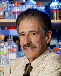
The David Graham Lab studies the consequences of HIV interactions with the immune system, the resulting pathogenesis and how to sabotage these interactions. We apply advanced technologies like mass spectrometry to dissect processes at the molecular level. We are also actively involved in cardiovascular research and studies the ways proteins are organized into functional units in different cell types of the heart. Major projects in our lab are organized into three major areas: (1) H/SIV pathogenesis and neuropathogenesis, (2) Cardiovascular disease, and (3) High technology development -
Zhu Lab
The Zhu lab is focused on characterizing the activities of large collection of proteins, building signaling networks for better understanding the mechanisms of biological processes, and identifying biomarkers in human diseases and cancers. More specifically, our group is interested in analyzing protein posttranslational modifications, and identifying important components involved in transcription networks and host-pathogen interactions on the proteomics level, and biomarkers in human IBD diseases.
-
William B. Guggino Lab
Work in the William B. Guggino Lab focuses on the structure of the cystic fibrosis transmembrane conductance regulator (CFTR) and water channels; the molecular structure of transport proteins in epithelial cell membranes; and gene therapies to treat cystic fibrosis (CF) patients. We are also working to identify CF’s specific defect in chloride channel regulation. One recent study showed that insulin-like growth factor 1 (IGF-1) enhances the protein expression of CFTR. -
Wei Dong Gao Lab
Work in the Wei Dong Gao Lab primarily focuses on heart failure and defining molecular and cellular mechanisms of contractile dysfunction. We use molecular biology and proteomic techniques to investigate the changes that myofilament proteins undergo during heart failure and under drug therapy. We're working to determine the molecular nature of nitroxyl (HNO) modification of tropomyosin. -
Landon King Lab
The Landon King Lab studies aquaporins water-specific membrane channel proteins. We hope to understand how these proteins contribute to water homeostasis in the respiratory tract and how their expression or function may be altered in disease states.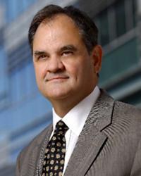
-
Krummey Lab
The Krummey Lab is a part of the Department of Pathology at the Johns Hopkins School of Medicine.
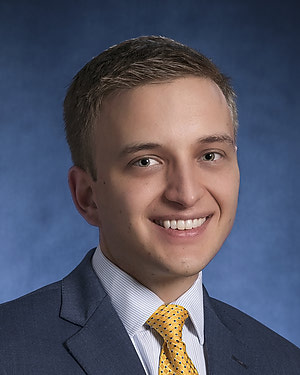
Our research prioritizes understanding the cellular mechanisms of alloimmunity, with a concentration on manipulating various cosignaling receptors and antigen recognition pathways to restrain the key lymphocytes principally involved in graft rejection. With the use of MHC tetramers, transgenic mouse models, and high-dimensional flow cytometry, we focus on mouse- and human-graft specific CD8+ T cells, CD4+ T cells, and B cells.
Transplantation is a life-saving procedure against a variety of diseases. Despite technical advances vastly improving early outcomes after transplant, long-term survival of transplanted organs has remained stagnant for the better part of three decades. A major cause of graft loss is immune-mediated rejection, which traditionally has be classified as acute or chronic based on its occurrence early or late after transplantation. Recently, this consensus has shifted to defining a graft rejection by its immunologic characteristics, either antibody-mediated or T cell-mediated (cellular rejection). This is because modern discoveries have identified the true major contributor to graft failures that occur many years after transplantation: not chronic rejection, but rather the cumulative impact of T cell-mediated acute rejection as a risk factor for later graft loss. Thus, original approaches to specifically prohibit and/or treat T cell-mediated acute rejection are of major significance for improving post-transplant outcomes.
HLA compatibility has also proven to be paramount for graft rejection. Originally, this was believed to be at the cellular level, then the single HLA protein level, and now at the epitope or molecular mismatch level. Specifically, HLA class II epitope-level mismatch has been identified as a risk factor for graft rejection, and multiple studies have identified specific epitopes within HLA class II peptides that are thought to be highly pathogenic. Few techniques directly measure antibody responses against specific regions of HLA proteins, but such measurements could provide both new information about the strength and character of alloimmunity and serve as an important new tool to study allogeneic B cells and antibody-secreting cells.
-
Mass Spectrometry Core
The Mass Spectrometry Core identifies and quantifies proteins that change expression in well-characterized protein fractions from cancerous cells or tissues. This includes identifying and quantifying changes in binding partners and post-translational modifications. Column chromatography and gel electrophoresis-based one and two-dimensional separations of protein complexes coupled to mass spectrometry are used. Techniques such as difference gel electrophoresis (DIGE), isobaric tag for relative and absolute quantitation (iTRAQ) and 18O-labeling as well as non-labeling methods (MudPit, multi-dimensional protein identification technology) are available for quantifying relative differences in protein expression and post-translational modifications. We developed methods to detect post-translational modifications such as LCMS methods to accurately determine the intact mass of proteins, selective fluorescent labeling of S-nitrosothiols (S-FLOS) to detect nitrosated cysteines in proteins, and ion mapping methods to map post-translational modifications that produce a signature mass or mass difference when the modified peptide is fragmented. -
Tom Woolf Lab
The Tom Woolf Lab studies the quarter of the genome devoted to membrane proteins. This rapidly growing branch of bioinformatics, which includes computational biophysics, represents the main research direction of our group. We aim to provide insight into critical issues for membrane systems. In pursuit of these goals, we use extensive computer calculations to build an understanding of the relations between microscopic motions and the world of experimental measurements. Our calculations use our own Beowulf computer cluster as well as national supercomputer centers. An especially strong focus has been on the computed motions of proteins and all-atom models of the lipid bilayers that mediate their influence. To compute these motions, we use the molecular dynamics program CHARMM. We hope to use our understanding of the molecular motions for the prediction of membrane protein structures using new computational methods. -
The Sun Laboratory
The nervous system has extremely complex RNA processing regulation. Dysfunction of RNA metabolism has emerged to play crucial roles in multiple neurological diseases. Mutations and pathologies of several RNA-binding proteins are found to be associated with neurodegeneration in both amyotrophic lateral sclerosis (ALS) and frontotemporal dementia (FTD). An alternative RNA-mediated toxicity arises from microsatellite repeat instability in the human genome. The expanded repeat-containing RNAs could potentially induce neuron toxicity by disrupting protein and RNA homeostasis through various mechanisms. The Sun Lab is interested in deciphering the RNA processing pathways altered by the ALS-causative mutants to uncover the mechanisms of toxicity and molecular basis of cell type-selective vulnerability. Another major focus of the group is to identify small molecule and genetic inhibitors of neuron toxic factors using various high-throughput screening platforms. Finally, we are also highly interested in developing novel CRISPR technique-based therapeutic strategies. We seek to translate the mechanistic findings at molecular level to therapeutic target development to advance treatment options against neurodegenerative diseases. -
O'Rourke Lab
The O’Rourke Lab uses an integrated approach to study the biophysics and physiology of cardiac cells in normal and diseased states. Research in our lab has incorporated mitochondrial energetics, Ca2+ dynamics, and electrophysiology to provide tools for studying how defective function of one component of the cell can lead to catastrophic effects on whole cell and whole organ function. By understanding the links between Ca2+, electrical excitability and energy production, we hope to understand the cellular basis of cardiac arrhythmias, ischemia-reperfusion injury, and sudden death. We use state-of-the-art techniques, including single-channel and whole-cell patch clamp, microfluorimetry, conventional and two-photon fluorescence imaging, and molecular biology to study the structure and function of single proteins to the intact muscle. Experimental results are compared with simulations of computational models in order to understand the findings in the context of the system as a whole. Ongoing studies in our lab are focused on identifying the specific molecular targets modified by oxidative or ischemic stress and how they affect mitochondrial and whole heart function. The motivation for all of the work is to understand • how the molecular details of the heart cell work together to maintain function and • how the synchronization of the parts can go wrong Rational strategies can then be devised to correct dysfunction during the progression of disease through a comprehensive understanding of basic mechanisms. Brian O’Rourke, PhD, is a professor in the Division of Cardiology and Vice Chair of Basic and Translational Research, Department of Medicine, at the Johns Hopkins University.