Research Lab Results
-
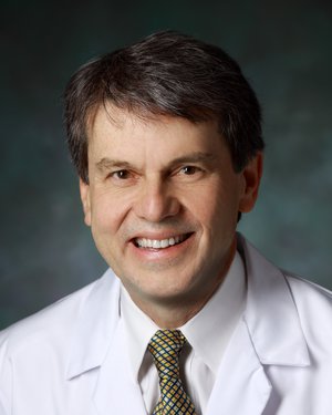
Wojciech Zbijewski Lab
Research in the Wojciech Zbijewski Lab — a component of the Imaging for Surgery, Therapy and Radiology (I-STAR) Lab — focuses on system modeling techniques to optimize the x-ray CT imaging chain. We’re specifically interested in: 1) using numerical models to improve the task-based optimization of image quality; 2) exploring advanced modeling of physics in statistical reconstruction; 3) using accelerated Monte Carlo methods in CT imaging; and 4) conducting experimental validation of such approaches and applying them to the development of new imaging methods.
-
Venu Raman Research Lab
The Raman laboratory is within the Division of Cancer Imaging Research in the Department of Radiology and Radiological Science. The focus of the laboratory is bench-to-bed side cancer research. We integrate molecular and cellular biology, developmental biology, cancer biology, molecular imaging techniques to study cancer formation and progression. Many of the projects in the lab investigate dysregulated genes in cancer and the translatability of this information to a clinical setting. One such project is to functionally decipher the role of a RNA helicase gene, DDX3, in the biogenesis of multiple cancer types such as breast, lung, brain, sarcoma, colorectal and prostate. Additionally, using a rational drug design approach, a small molecule inhibitor of DDX3 (RK-33) was synthesized and its potential for clinical translation is being investigated.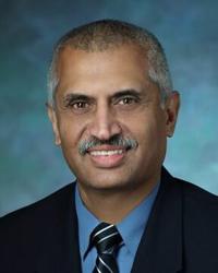
-
Kristine Glunde Lab
The Glunde lab is within the Division of Cancer Imaging Research in the Department of Radiology and Radiological Science. The lab is developing mass spectrometry imaging as part of multimodal molecular imaging workflows to image and elucidate hypoxia-driven signaling pathways in breast cancer. They are working to further unravel the molecular basis of the aberrant choline phospholipid metabolism in cancer. The Glunde lab is developing novel optical imaging agents for multi-scale molecular imaging of lysosomes in breast tumors and discovering structural changes in Collagen I matrices and their role in breast cancer and metastasis. -
Lima Lab
The Lima Lab’s research is concentrated on the development and application of imaging and technology to address scientific and clinical problems involving the heart and vascular system. Specifically, our research is focused on developing magnetic resonance imaging (MRI) contrast techniques to investigate microvascular function in patients and experimental animals with myocardial infarction; functional reserve secondary to dobutamine stimulation and myocardial viability assessed by sodium imaging; and cardiac MRI and computed tomography (CT) program development of techniques to characterize atherosclerosis in humans with cardiovascular or cerebrovascular disease. Current projects include: • The Coronary Artery Risk Development in Young Adults (CARDIA) Study • The MESA (Multi-Ethnic Study of Atherosclerosis) Study • The Coronary Artery Evaluation using 64-row Multidetector Computed Tomography Angiography (CORE64) Study Joao Lima, MD, is a professor of medicine, radiology and epidemiology at the Johns Hopkins School of Medicine. -
Neuromodulation and Advanced Therapies Center
We investigate the brain networks and neurotransmitters involved in symptoms of movement disorders, such as Parkinson's disease, and the mechanisms by which modulating these networks through electrical stimulation affects these symptoms. We are particularly interested in the mechanisms through which neuromodulation therapies like deep brain stimulation affect non-motor brain functions, such as cognitive function and mood. We use imaging of specific neurotransmitters, such as acetylcholine and dopamine, to understand the changes in brain chemistry associated with the clinical effects of deep brain stimulation and to predict which patients are likely to have changes in non-motor symptoms following DBS. Through collaborations with our neurosurgery colleagues, we explore brain function by making recordings during DBS surgery during motor and non-motor tasks. Dr. Mills collaborates with researchers in the Department of Neurosurgery, the Division of Geriatric and Neuropsychiatry in the Department of Psychiatry and Behavioral Sciences and in the Division of Nuclear Medicine within the Department of Radiology to translate neuroimaging and neurophysiology findings into clinical applications.
-
Quantitative Imaging Technologies
Research in the Quantitative Imaging Technologies lab — a component of the Imaging for Surgery, Therapy and Radiology (I-STAR) Lab — focuses on novel technologies to derive accurate structural and physiological measurements from medical images. Our team works on optimization of imaging systems and algorithms to support a variety of quantitative applications, with recent focus on orthopedics and bone health. For example, we have developed an ultra-high resolution imaging chain for an orthopedic CT system to enable in-vivo measurements of bone microstructure. Our interests also include automated methods to extract quantitative information from images, including anatomical and micro-structural measurements, and shape analysis.
-
Interventional Cardiology Research Group
Our group is interested in a broad array of clinical and translational investigations spanning the evaluation of basic pathophysiology in patients undergoing cardiac procedures, development and evaluation of new therapeutic strategies, and improving patient selection and outcomes following interventional procedures. We are comprised of a core group of faculty and dedicated research nurses as well as fellows, residents, and students. Projects range from investigator-initiated single-center observational studies to industry-sponsored multicenter phase 3 randomized controlled trials. We have established a database of all patients who have undergone TAVR at Johns Hopkins, which is providing the basis for several retrospective analyses and will serve as the foundation for future studies of TAVR. We are also engaged in collaborative projects with other groups from the Department of Medicine and other Departments including Cardiac Surgery, Anesthesiology, Radiology, Psychiatry, and Biomedical Engineering. Members of our group are actively involved with the Johns Hopkins Center for Bioengineering Innovation and Design (CBID) in the development of novel minimally-invasive cardiovascular devices.
-
Marie-France Penet Lab
The Penet lab is within the Division of Cancer Imaging Research in the Department of Radiology and Radiological Science. The lab research focuses on using multimodal imaging techniques to better understand the microenvironment and improve cancer early detection, especially in ovarian cancer. By combining MRI, MRS and optical imaging, we are studying the tumor microenvironment to understand the role of hypoxia, tumor vascularization, macromolecular transport and tumor metabolism in tumor progression, metastasis and ascites formation in orthotopic models of cancer. We also are studying the role of tumor-associated macrophages in tumor progression. -
Brain Health Program
The Brain Health Program is a multidisciplinary team of faculty from the departments of neurology, psychiatry, epidemiology, and radiology lead by Leah Rubin and Jennifer Coughlin. In the hope of revealing new directions for therapies, the group studies molecular biomarkers identified from tissue and brain imaging that are associated with memory problems related to HIV infection, aging, dementia, mental illness and traumatic brain injury. The team seeks to advance policies and practices to optimize brain health in vulnerable populations while destigmatizing these brain disorders. Current and future projects include research on: the roles of the stress response, glucocorticoids, and inflammation in conditions that affect memory and the related factors that make people protected or or vulnerable to memory decline; new mobile apps that use iPads to improve our detection of memory deficits; clinical trials looking at short-term effects of low dose hydrocortisone and randomized to 28 days of treatment; imaging brain injury and repair in NFL players to guide players and the game; and the role of inflammation in memory deterioration in healthy aging, patients with HIV, and other neurodegenerative conditions. -
The Pathak Lab
The Pathak lab is within the Division of Cancer Imaging Research in the Department of Radiology and Radiological Science. We develop novel imaging methods, computational models and visualization tools to ‘make visible’ critical aspects of cancer, stroke and neurobiology. Our research broadly encompasses the following areas: Functional and Molecular Imaging; Clinical Biomarker Development; Image-based Systems Biology and Visualization and Computational Tools. We are dedicated to mentoring the next generation of imagers, biomedical engineers and visualizers. Additional information can be found at www.pathaklab.org or by emailing Dr. Pathak.