The Johns Hopkins Applied Imaging Mass Spectrometry – AIMS Core/Service Center

The Johns Hopkins Applied Imaging Mass Spectrometry (AIMS) Core/Service Center provides rapid matrix-assisted laser desorption/ionization (MALDI) imaging at high spatial resolution, which includes sample preparation, on-tissue digests and derivatizations, and data analysis.
Spatially resolved MALDI imaging measurements are directly taken from a frozen or formalin-fixed paraffin-embedded (FFPE) tissue section without destroying it. MALDI imaging combines mass spectrometric analyses of biomolecules with simultaneous histological evaluation to analyze intact proteins, peptides and tryptic peptides (on-tissue tryptic digest), N-glycans (on-tissue PNGase digest), lipids, metabolites and drug molecules in a spatially resolved manner. The Johns Hopkins AIMS Core provides comprehensive service in MALDI tissue imaging to investigators at Johns Hopkins and outside.
Equipment
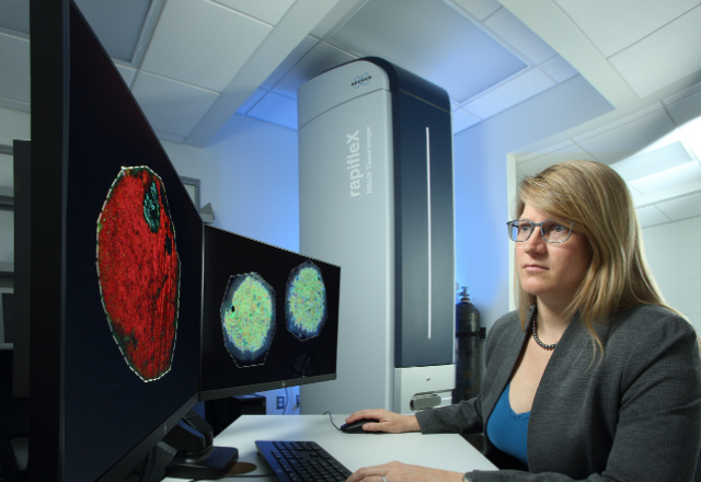
- Bruker Rapiflex MALDI TOF/TOF instrument for high-throughput MALDI imaging
- HTX M5 sprayer for accurate robotic spraying of enzymes and matrices
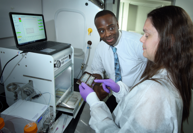
- Leica cryostat for MALDI imaging-compatible cryosectioning
- Slide scanner for histology and immunohistochemistry co-registered with MALDI imaging
- High-end workstation with SCiLS lab for data analysis
Services
- MALDI imaging compatible cryosectioning in gelatin and other MALDI compatible cryomedia
- MALDI imaging sample preparation of formalin-fixed, paraffin-embedded tissue sections
- On-tissue digestions including for tryptic peptides and glycans
- On-tissue derivatizations for metabolite imaging
- Robotic matrix application with HTX M5 sprayer
- High-throughput multiplexed MALDI imaging of up to 5,000 biomolecules at once
- High spatial resolution MALDI imaging of 20 micron pixel size or better
- MALDI imaging covering maximum area of regular microscopy slide (75 millimeters by 25 millimeters)
- Customized development of MALDI imaging protocols
- Targeted MALDI imaging of drugs, drug metabolites, imaging agents, contrast agents or other agents
- Discovery MALDI imaging of metabolites, lipids, peptides, intact proteins, tryptic peptides and glycans
- On tissue MS/MS for analyte identification in imaging or profiling mode
- Data analysis: segmentation analysis, pathology-guided analysis and statistical analysis with dedicated software package (SCiLS Lab)
Leadership
Kristine Glunde, M.S., Ph.D.
Director
Email: [email protected]
Telephone: 410-614-2705 (office), 410-614-7959 (AIMS Core)
Contact Information
For more information, please contact:
Dr. Kristine Glunde
The Russell H. Morgan Department of Radiology and Radiological Science
The Johns Hopkins University School of Medicine
Office: Traylor Building, Room 203, Rutland Avenue, Baltimore, Maryland 21205
AIMS Core: Cancer Research Building II, Room LB03D/E, Baltimore, Maryland 21231