What to Expect at Your Appointment at the Pediatric and Congenital Heart Center
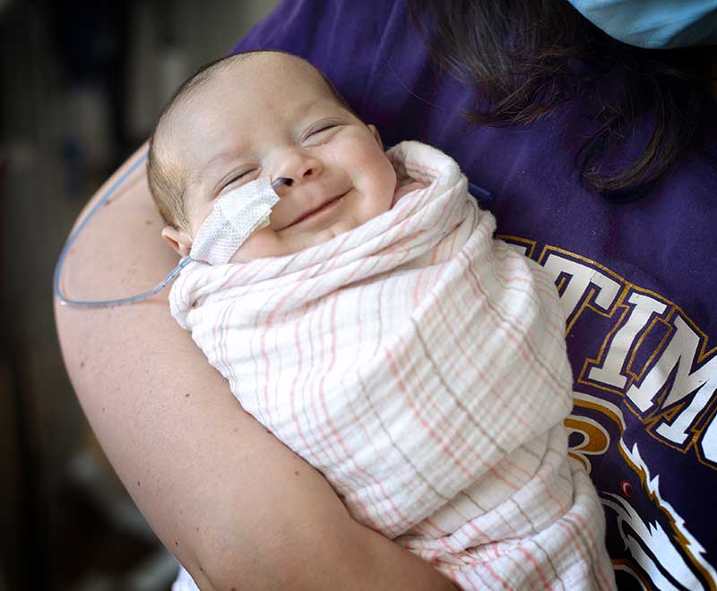
Welcome to the Pediatric and Congenital Heart Center, and thank you for entrusting us with your child’s heart. We look forward to providing each infant, child or teen with a tailored plan, comprising the most effective diagnostic, medical and surgical approaches. Whether your child has an congenital heart problem or is experiencing acute illness, you and your family are in the right place.
Learn what to expect during the following tests and procedures:
Imaging
Children’s heart treatment: Seeing the challenge clearly
The advanced imaging technology available through our center helps us see details of the smallest, most intricate structures in your child’s heart, from fetuses and newborns to older children and adults with congenital heart conditions. The technologists who create the images share their captured images with surgeons and other doctors to help plan the most effective treatment.
Imaging Your Child’s Heart: What to Expect
Imaging tests such as computed tomography (CT) or magnetic resonance imaging (MRI), echocardiogram, or electrophysiology (EKG) can show structural problems in the heart. They are performed by a technologist, who takes the images to the cardiologist. The cardiologist discusses the findings with you. Results of your child’s tests will appear on MyChart, often the same day.
MRI and CT are two ways to see inside your child’s heart — painlessly
These techniques can help the cardiologist visualize arteries of the lung and heart, as well as structures in and around the heart itself. The scans themselves do not hurt. The CT equipment we use delivers a very low level of radiation. They require your child stay still and may or may not involve an injection of a contrast agent for more accurate pictures.
3-D Printing and Augmented Reality
Complex pediatric heart conditions require exceptional imaging. Our center has access to advanced imaging that reveals lifelike details of your child’s heart and blood vessels, using some of the same technology developed for computer games. We can even print these images in three dimensions to help us plan treatment.
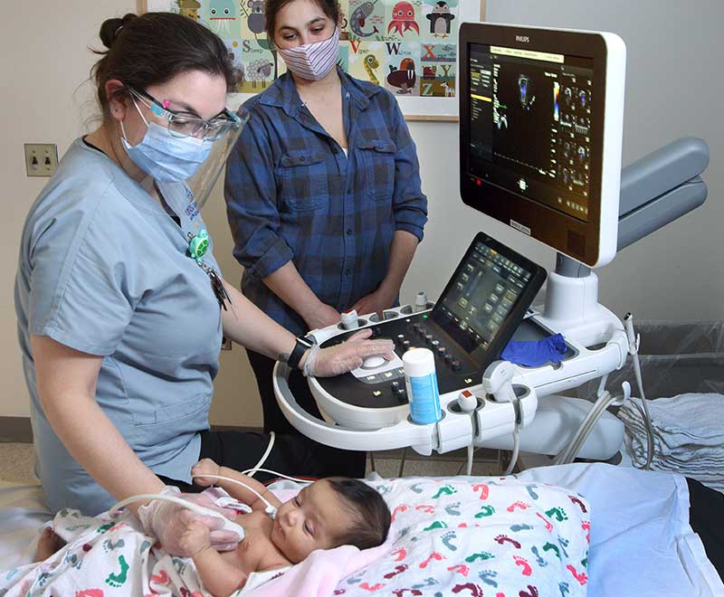
Echocardiography can reveal a lot about your child’s heart
Echocardiography is a type of sonogram for the heart that can identify problems in its chambers and the passageways between them (valves). It uses painless sound waves to create an image of the heart and its structures.
Echocardiograms: from Outside and Inside
Echocardiograms provide a great deal of information to your child’s cardiologist without using radiation. Echocardiography images can be captured from outside of the body, using a transducer, or from inside the body (transesophageal echocardiogram, or TEE.)
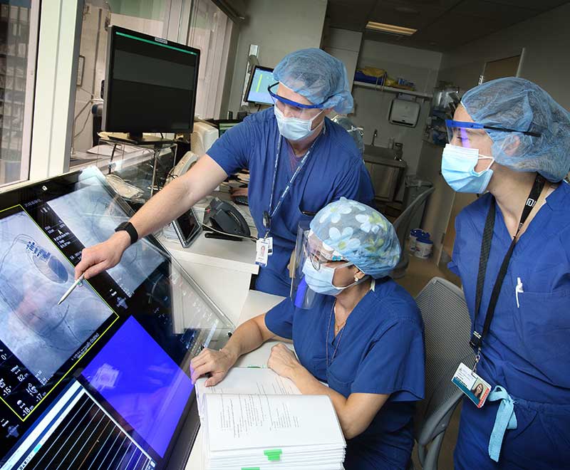
When our surgeons are considering a catheterization or surgery, we sit down as a group and go over the images we've collected, one on one. We prepare the surgeon for the procedure with 3-D imaging and prints.
Melanie Nies, M.D.
Electrophysiology
Examining Your Child for Arrhythmias
An electrocardiogram (ECG) or other electrophysiology test records electrical activity in your child’s heart and help identify abnormal heart rhythms (arrhythmias). Some electrophysiology heart rhythm tests can be conducted at our center, and others track your child’s arrhythmia from home using monitors or implantable loop recorders.
Radiofrequency ablation (RFA) can help correct abnormal heart rhythms
Your child’s cardiologist might recommend catheter-guided radiofrequency ablation, or RFA, to treat abnormal heart rhythms (arrhythmias). This procedure targets heart cells that are sending the wrong electrical signal to the heart muscle.
RFA: Answering Your Questions
Our doctors meet with you and go over what will occur, step by step so you can visualize the process. You can see the instruments and the devices the doctor will use to repair your child’s heart, and get answers to your questions about risks and benefits, anesthesia and other aspects of treatment.
We are committed to each child throughout every procedure and throughout their lives. Whenever we meet with a new family, we stress that we are not going to do anything that isn’t necessary. Our job is to keep your child safe, no matter what.
Hannah Fraint, M.D.
Cardiac Catheterization
Catheterization offers solutions without surgery
Catheterization techniques may be able to help doctors repair some heart problems without surgery. Catheterization can help address inborn heart malformations, pulmonary artery problems, valve replacement, and holes in the heart or vessels.
Heart Catheterization: What Happens
In performing catheterization, the doctor inserts a very thin, flexible tube through a blood vessel, using a guide wire, and eases the catheter through the blood vessels to the heart, where the doctor can repair malformations or replace implanted devices such as valves.
Heart Failure and Pediatric Heart Transplant
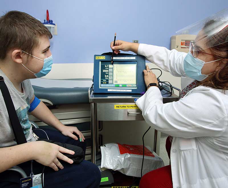
Ventricular Assist Devices (VAD)
For children affected by heart failure, a ventricular assist device, or VAD, can work as a mechanical pump to help a weakened or damaged heart circulate blood through the body. By itself, a VAD is not a permanent treatment for heart failure, but is considered a “bridge” to improve children’s quality of life while they await a heart transplant.
VADs: Options for Treatment
There are different types of VADs; infants’ models are often outside of the body while older children and teens may have an implanted VAD.
Covering Every Aspect of Recovery from Heart Failure
From surgery preparation to recovery, we will make sure you will understand what is happening when, and what you can expect. Collaboration between pediatric cardiologists, specialists in children’s critical care and anesthesia, clinical nurses, cardiac child life specialists and social workers gives your child the best chance for a healthy future.
Continuous Care after Pediatric Heart Failure
Children and teens whose heart failure can be stabilized with treatment or surgery will require ongoing care, with regular outpatient catheterization procedures, further biopsies and possible surgery. A successful approach can mean a full life for your daughter or son, with an eventual return to physical activity, school, normal socializing and relatively few limitations.

Surgery and Intensive Care
If Your Child Needs Heart Surgery
From the simplest and most common heart issues to the most rare and complex cases, we offer surgical skill, well-coordinated teamwork, and compassionate care for you child. Collaboration with pediatric experts in otolaryngology, critical care, genetics and other specialties ensures a highly skilled and individualized approach to your child’s care.
The Pediatric Cardiac ICU is unique
Specialized cardiac critical care of children with heart problems, as delivered through our PCICU, has many advantages, including shorter times on ventilators and improved continuity and coordination among the specialists caring for your child.
Learn more about preparing your child for surgery at Johns Hopkins Children’s Center.
Dedicated Pediatric Cardiac Unit
Our innovative pediatric cardiac unit (PCU) is designed to strengthen care and reduce length of stay and readmissions for patients discharged from the pediatric cardiac intensive care unit. Multidisciplinary services on the PCU include pulmonology, Child Life, nutrition, feeding specialists, physical and occupational therapy, respiratory therapy and social work. While on the unit, patients are monitored with state-of-the-art telemetry to speed recovery and safely simulate home activities.
Fetal Cardiology
If your obstetrician has identified a possible heart problem in your unborn baby, our skilled, multidisciplinary team is ready to help, regardless of the stage of your pregnancy. For non-cardiac issues, our team provides expert cardiology support, including monitoring during fetal surgery performed by our colleagues in maternal-fetal medicine. After delivery, our expertise ensures the best possible care for babies in the neonatal intensive care unit (NICU).
Supporting Your Family as Your Child Receives Care