Patient Story
Detached Lens: Ollie's Story
Ollie Cantos was walking home from work one evening in May 2019. A block from his office, he approached a crosswalk. On the side street, a truck waited for him to cross. A jeep with an impatient driver, however, veered around the truck into the intersection and hit Cantos.
He was treated at a local hospital for injuries that included a fractured foot bone. He remembers thinking, “I’m hurt, but I’ll get better.” What he didn’t know was that the force of the collision had caused the lens in his right eye to partially detach. Within several months, it would detach completely.
The eye’s lens is a transparent, disk-shaped structure that focuses light onto the retina, allowing us to see clearly. Located behind the pupil, it is held in place by fine ligaments, which tore on impact in Cantos’s eye.
For Cantos, who had been born with retinopathy of prematurity (ROP), the situation was dire. As its name suggests, ROP affects premature infants. It occurs when abnormal blood vessels grow throughout the retina — the tissue that lines the back of the eye — in response to incomplete development of the retinal vasculature. The blood vessels are fragile and can leak, scarring the retina and causing it to detach, which in turn can lead to visual impairment and blindness.
In Cantos’s case, he never had vision in his left eye, and at the time of the accident, vision in his right eye was limited. Still, with the aid of magnification devices, text-to-speech technology and a cane, he could read and maneuver around his home and community. He was also raising three boys, triplets he had adopted who had ROP and were totally blind. Then there was his work: Cantos had a rewarding career as an attorney at the U.S. Department of Education.
Now, however, even as he healed from the other injuries sustained in the accident, Cantos began to realize what he calls his worst nightmare. At first, he noticed a sort of split screen, where everything on one part of his field of vision had a yellowish tinge. But when the lens detached completely, it began floating around freely in his eye and in his field of vision.
“If you can imagine looking at an amoeba under a microscope and it swimming around and having tentacles, that's what I saw with my eye open or closed, 24/7,” Cantos recalls. “It nearly made me crazy. I couldn’t look at anything.”
At work, Cantos found it impossible to stay focused. “I would try to move my head to the side to optimize it [the lens] being out of the way,” he says. “Then suddenly, when I was talking to people, it would move in the way again.”
Cantos’s ophthalmologist diagnosed detached lens. It wasn’t harming his health, the doctor said, and if they tried to remove the lens, there was a very real risk he would lose what little vision he had.
‘Get Used to It’
For people with no complicating factors, the success rate of surgery to repair a detached lens is about 98%. But ROP had caused scar tissue to develop in the back of Cantos’s eye, and the retina was very thin and liable to detach. If anything went wrong, he could become completely blind. “Maybe you’ll get used to it over time,” the doctor told him.
But Cantos could not get used to it. Living with the floating lens was tantamount to torture. Even sleep was elusive. When he closed his eyes, if there was even a hint of light in the room, he could see the floating lens. Cantos was accustomed to using modifications to enhance his ability to see, including a bright background on his phone. Now the bright background only illuminated the floating lens more. Increasingly, he found himself crying.
He again pleaded with the doctor to do something, and again, the doctor said that while he could remove the lens, Cantos might go blind. And the ophthalmologist had no experience with such a delicate operation. He suggested that Cantos contact the National Eye Institute (NEI).
Part of the National Institutes of Health, NEI conducts and supports research, training and other programs regarding blinding eye diseases and visual disorders. None of the doctors there had ever removed a lens from someone with ROP, and the risk, they said, was extraordinarily high. The NEI team suggested that Cantos go to Wilmer Eye Institute, Johns Hopkins Medicine, which deals with all kinds of challenging conditions and specializes in complex cases. They even gave him a name: Dr. Fernando Arevalo.
In Expert Hands
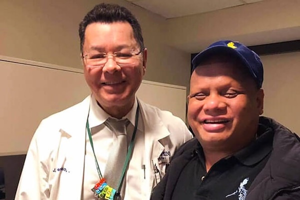
J. Fernando Arevalo, M.D., Ph.D., chairman of ophthalmology at Johns Hopkins Bayview Medical Center, and the Edmund F. and Virginia Ball Professor of Ophthalmology at the Wilmer Eye Institute, specializes in vitreoretinal diseases. A gifted surgeon, he has performed thousands of retinal detachment operations and conducted research that has led to advances in the treatment of complex retinal detachments and other conditions.
Arevalo told Cantos that while he had never removed a lens in a patient with ROP, he had performed surgery on patients with ROP, and he had performed more than 10,000 vitrectomies — surgeries to address a variety of problems, including retinal detachments and detached lenses.
"I thought, well, yes, he has all these issues in the back of the eye that we cannot change,” Arevalo recalls, “but if we don’t take care of this lens floating around in the back of the eye, basically creating a big shadow over his already poor vision, he won’t be able to do his normal activities at all. He’s essentially going to be blind anyway.”
“He was so unbelievably kind,” Cantos says of Arevalo. “He told me that there is a risk and that I needed to understand that if it doesn't go well, I could be totally blind because of complications like bleeding. I told him, ‘I don't care what the risk is. I need to have the surgery, but I need to have it done by the best doctor.”
In February, after more than nine months of agony, Cantos had surgery at the Wilmer Eye Institute to remove the lens. Afterward, a bandage was over his eye. Arevalo had placed air in the eye to help the retina stay in its proper position during healing and to maintain good eye pressure, and initially Cantos could see what looked like little gray bubbles. Stimulation of the nerves in the eye temporarily caused Cantos to see a very bright light, even with the eye closed and bandaged. But the first thing Cantos noticed — before the light, the bubbles and the bandage – was that the lens, the thing that had tormented him all those months, was gone.
Forever Grateful
Before the surgery, Arevalo offered to implant a replacement lens to maximize Cantos’s remaining vision, but he said it would further risk damage to the fragile tissues in the eye. Cantos decided that while having the surgery was vital to his quality of life, he didn’t want the additional risk. Although he did lose some vision with the removal of his natural lens, Cantos is adamant that the surgery was the right decision for him.
“I'm grateful for what I have,” he says. “I have my family. I have a job that I love. I have people at work who have been incredible throughout all of this. And I’m forever grateful to Dr. Arevalo, because life is still a billion, trillion, quadrillion times better. Dr. Arevalo and the whole team at Wilmer make life better for countless numbers of us. Our lives are transformed.”
Treatment Wilmer Eye Institute