Spinal Angiogram
What You Need to Know
- Spinal angiography uses dye and X-rays to help doctors see the veins and arteries around the spinal cord.
- The dye is given through a catheter inserted into an artery in the groin.
- This procedure can help diagnose blood vessel malformations, blood clots and other problems, as well as help doctors plan treatments for these conditions.
- A spinal angiogram is most often performed under light sedation, and most patients can return home the same day.
What is spinal angiography?
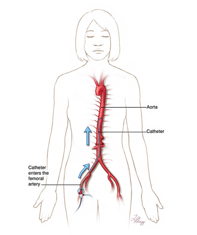
A spinal angiogram is a medical procedure that uses dye and low-dose X-rays to get a detailed look at the blood vessels surrounding the spine and spinal cord. The test is performed by specially trained doctors called neuro-angiographers.
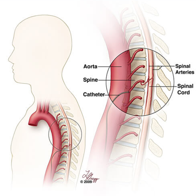
Dye is slowly injected into an artery feeding the spine or spinal cord via a catheter inserted in the groin. As the dye fills the arteries and veins, it shows up on an X-ray, providing a clear picture that allows doctors to identify problems such as:
- Narrowing or blockages in the blood vessels
- Abnormal shape or bulging of the vessels
- Blood vessel malformations
- Tears that cause bleeding
- Hardening and other blood vessel diseases
- Tumors of the spine or spinal cord
Who might need a spinal angiogram?
Your doctor may recommend spinal angiography if he or she suspects you have a condition affecting blood vessels around the spine or spinal cord. Those may include:
- Spinal cord vascular malformations, such as dural arteriovenous fistulas and arteriovenous malformations
- Certain types of spinal cord stroke: An artery surrounding the spinal cord can become narrowed due to blood vessel disease. A piece of plaque or a blood clot can block the diseased artery and cut off blood supply to part of the spinal cord.
- Spinal venous thrombosis, a blockage in a vein near the spinal cord.
- Spine and spinal cord tumors
Spinal angiography is sometimes used in situations when an MRI cannot provide a clear picture. Interventional neuroradiologists may also use spinal angiography to plan treatments for the above conditions, such as blocking blood supply to a vascular mass (embolization). Certain treatments can be performed at the same time as spinal angiography, with X-rays providing guidance.
Preparing for a Spinal Angiogram
Your doctor will explain the procedure to you, and will ask if you have questions. You will need to provide a list of medications you are taking, including all prescribed and over-the-counter drugs and all herbs, vitamins and supplements.
Tell your doctor and the people performing the test if you:
- Have allergies
- Have ever had a reaction to any contrast dye
- Have any problems with your kidneys
- Are pregnant or might be pregnant
- Have any bleeding or blood clotting disorders
- Take blood thinner medications
In preparation for a spinal angiogram, your doctor may ask you to:
- Avoid eating anything for a certain time before the procedure. The fasting period may be a few hours or overnight.
- Have a blood test to see how long it takes your blood to clot. Other blood tests may be done as well.
Based on your medical condition, your physician or other health care provider may give you other instructions on what to do before the procedure.
What happens during spinal angiography?
The procedure takes place in a room specifically designed for angiography tests on the brain and spine. The angiography suite is equipped with a table surrounded by rotating X-ray machines and monitors.
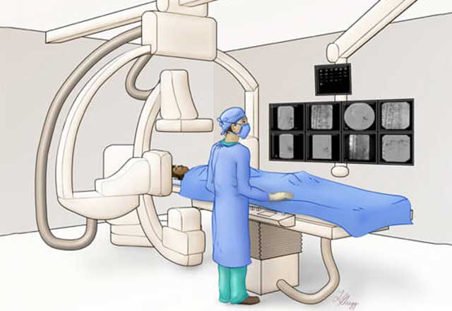
Spinal angiography typically involves the following steps:
- Once you change into a gown and are comfortably positioned on the table, the angiographer will give you medicine by vein that will help you relax.
- Next, you will get a shot of numbing medicine in the groin area, which may cause a brief, stinging sensation.
- When the area is numb, the angiographer will insert a catheter (a narrow, flexible tube) into one of the arteries in your groin. This vessel connects to the vessels of the spine and spinal cord.
- After the angiographer positions the catheter near the suspected problem areas, a small amount of contrast dye will be injected. You may feel a sensation of warmth for a few seconds as the dye moves through your blood vessels.
- While the dye flows through your veins and arteries, the angiographer takes X-ray pictures of the blood vessels. Several injections of dye are necessary to get pictures of all the small blood vessels of the spinal cord.
- The pictures are recorded and displayed on screens that the angiographers watch as they are performing the procedure.
- At the end of the procedure, the catheter is removed from the groin and pressure is applied to the incision for 15–20 minutes to control bleeding. Alternatively, a small plug is used to close the puncture site.
- You will move to a recovery room, where a nurse will monitor you. Meanwhile, the doctors closely review the recorded images, and you should get a result before you leave.
Recovery After a Spinal Angiogram
The most important thing after an angiogram is to prevent bleeding at the site where the catheter was inserted. It takes time for this incision to seal. If the angiographers have closed the incision with a plug, you will need to lie down and keep your leg straight for up to three hours to make sure the incision does not bleed. If a plug was not used, this time could be longer ― up to six hours. In rare instances, you may need to spend the night at the hospital. You may get medicine to help you stay comfortable while you lie flat.
After the recovery period, you can go home or go back to your hospital room. If you are leaving the care facility, make sure you have someone to drive you home.
Once you are home, drink plenty of water and other fluids to help you get rid of the contrast dye in your body. You can go back to eating normally. Follow your doctor’s instructions on when you can resume bathing, exercise and other strenuous activities. Most people can return to work a day or two after the procedure.
A small bruise at the place where the incision was made is normal. You might notice slight seeping or an occasional drop of blood.
If the spinal angiogram shows a problem with the blood vessels surrounding your spinal cord, your doctor will follow up with you and work with you on a treatment plan.
Risks and Complications of Spinal Angiography
Diagnostic spinal angiography is very safe, especially when performed by experienced neuro-angiographers. Newer techniques have dramatically lessened the chance of a spinal stroke ― the most serious but very rare complication of a spinal angiogram. The angiographer will review the risks and benefits of angiography and answer all your questions before the procedure.
Contact your doctor if you experience:
- Any changes to your leg, such as unusual redness, heat or coolness, pain, numbness, tingling or weakness
- Fever or chills
- Speech or vision changes
- Dizziness
- Chest pain