Laser Interstitial Thermal Therapy (LiTT)
Laser interstitial thermal therapy, or LiTT, is a minimally invasive procedure that uses heat to destroy areas of abnormal cells, such as tumors or cells causing seizures.
Laser interstitial thermal therapy is sometimes called laser ablation surgery, stereotactic laser ablation or MRI-guided laser ablation.
What You Need to Know
- Laser interstitial thermal therapy (LiTT) is a minimally invasive procedure that is most commonly used to treat epilepsy, brain tumors and a condition called radiation necrosis.
- The treatment uses heat to ablate (destroy) cancer cells, areas of dead tissue or a brain lesion where seizures begin.
- The procedure uses MRI guidance and takes several hours, followed by one or two nights in the hospital.
- Compared with those who undergo traditional open surgery, people who have LiTT may experience less pain and have a faster recovery.
What is laser interstitial thermal therapy?
Laser interstitial thermal therapy is a procedure that destroys abnormal tissue, typically in the brain, using heat from laser beams guided by MRI. It is a minimally invasive surgical alternative to traditional open skull surgery (craniotomy).
A LiTT device features a probe equipped with special fibers, which is inserted through a small hole in the skull. Once the tip of the probe is positioned in the problem area using MRI guidance, a laser beam is directed through the probe to heat up the tip and damage the surrounding abnormal tissues, causing cell death. Special MRI sequences are used to measure the temperature of the surrounding brain, helping ensure there is no damage to nearby healthy structures.
Compared with craniotomy, LiTT may provide advantages such as less pain after the procedure, shorter recovery and fewer cognitive problems after treatment. The incision is usually about a half-inch long, patients tend to go home the next day, and they return to work more quickly than after a craniotomy.
Who may need the LiTT procedure?
To determine if LiTT is the right approach for your condition, your care team will request and review all medical records and tests, especially EEGs, brain images and neuropsychological tests. The doctors may recommend further lab work, biopsies and imaging to determine if LiTT is the most appropriate treatment for you. Sometimes, a biopsy may be performed at the same time as LiTT.
LiTT Procedure for Epilepsy
Some forms of epilepsy are caused by a lesion in the brain such as a scar or a malformation of brain tissue or blood vessels. These lesions can cause focal seizures, which may not be controlled with epilepsy medication. LiTT may be used to treat the problem lesions and reduce or eliminate the seizures they cause.
For example, one type of focal epilepsy called mesial temporal lobe epilepsy (MTLE), often treated with surgery to remove the lesion, may be treated with LiTT instead. Studies have shown that LiTT is nearly as successful as traditional craniotomies in addressing MTLE.
LiTT can also be used to treat focal epilepsy and other neurological problems caused by certain benign growths and malformations in the brain. LiTT can successfully remove growths such as:
- Periventricular nodular heterotopia – a malformation of gray matter cells in the brain
- Hypothalamic hamartoma – a benign growth on the hypothalamus
- Cavernous malformation – an abnormal collection of blood vessels
- Focal cortical dysplasia – a malformed area in the top layer of the brain
LiTT can be an alternative to disconnection procedures such as corpus callosotomy. However, when corpus callosotomy is needed, laser ablation can be used to sever the connection instead of cutting.
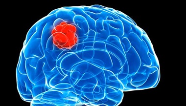
LiTT Procedure for Brain Tumors
MRI-guided laser ablation such as LiTT may make it easier for neurosurgeons to gain access to some tumors that are located close to sensitive structures or deep in the skull, where they would be hard to reach and remove safely through traditional open craniotomy.
In some cases, LiTT may not completely remove a brain tumor but may be able to shrink it or change the texture of the tumor tissue so it is easier to remove through a craniotomy. If a diagnosis is needed, biopsies can be performed during the same procedure as an ablation.
Smaller brain tumors
Smaller brain tumors (those less than 3 to 5 centimeters across) may be able to be treated with LiTT, including glioblastoma, a dangerously aggressive glioma tumor that frequently comes back and can be life-threatening. LiTT can also be used on less advanced gliomas and benign tumors such as a meningioma located in an area where it would be difficult to remove with surgery.
Palliative care
LiTT can be used to help shrink tumors as part of palliative care, which addresses symptoms and can improve the patient’s quality of life.
Brain metastases
LiTT can also be used to treat brain metastases (when cancer spreads to the brain from other parts of the body). LITT is considered safe and effective for brain metastases, especially those that are small or located deep in the brain.
LiTT for Radiation Necrosis
In 9% to 14% of cases of a person receiving stereotactic radiosurgery for brain cancer, that treatment may leave behind areas of scar tissue, damaged blood vessels and dead cells (necrosis). About half the time, radiation necrosis goes away on its own. But necrotic areas in the brain can, over time, cause symptoms due to swelling and irritation in the brain, including a higher risk of infection.
Radiation necrosis can be addressed with surgery, pure oxygen (hyperbaric) treatments, steroids, blood thinners and other drugs. For some patients, LiTT may be an effective alternative to surgery in removing the dead tissue.
Using LiTT for radiation necrosis was first reported by U.S. doctors in 2012. Since then, data show that about 75% of people treated with LiTT have had relief from radiation necrosis symptoms.
Other Uses of LiTT
Outside of the central nervous system, LiTT and similar laser ablation techniques have been used for treatment of tumors in the following areas:
- Lung and central airway
- Liver
- Prostate
- Bone
Research studies are exploring other uses of LiTT, including treatment of metastatic spine cancer, which is cancer from another part of the body that has spread (metastasized) to the spine.
Preparing for a LiTT Procedure
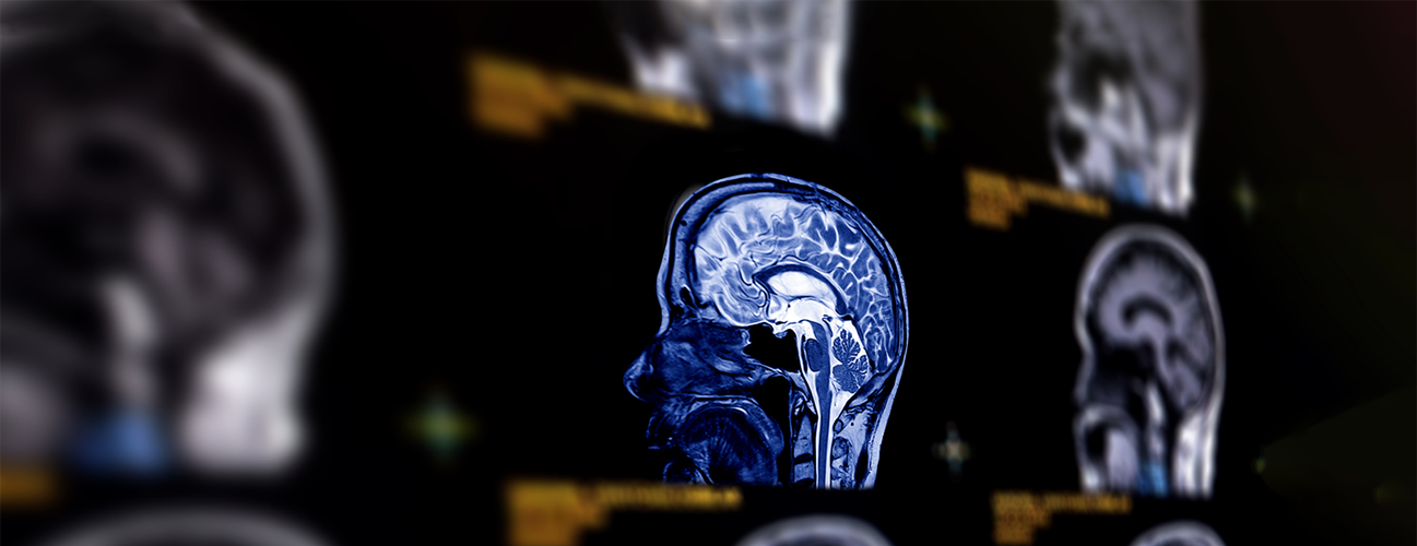
Before your procedure, while you are in the operating room, the doctors capture images of the area to be treated using MRI. These images are sent to the LiTT workstation, which creates “maps” targeting the spots where the lasers will be applied. Precise measurement and skilled guidance help protect healthy tissue from damage.
What happens during the LiTT procedure?
LiTT is typically performed in a surgical room equipped with an intraoperative MRI machine. You will be under general anesthesia and inside the MRI machine for most parts of the procedure, which takes about three hours.
- Once you are asleep under general anesthesia, a special frame may be attached to the scalp to hold the head very still.
- The surgeon makes a small hole in the skull at a location that makes it easier to access the spot for treatment.
- Using a grid-like map called stereotactic navigation, the surgeon directs a needle-like fiberoptic laser probe toward the area in the brain where the problem lesion is located. The probe placement is confirmed by the MRI images.
- Laser heat, between about 113 to 138 degrees Fahrenheit, is delivered to the tip of the probe to destroy the abnormal tissue. The heat is carefully monitored with MRI thermometry to avoid overheating the surrounding area. The tip of the applicator is cooled with saline or carbon dioxide, depending on the brand of LiTT machine used.
- Once the area has been treated, the neurosurgeon removes the probe and the incision is closed with one or two sutures (stitches).
Recovery After the LiTT Procedure
You will spend one or two nights in the hospital for observation. Your doctor may prescribe medicine to address swelling. Most people can begin to return to their normal activities in about a week — a much shorter time than after craniotomy. Complete recovery may take several weeks.
The LiTT procedure may need to be repeated, for instance, if a brain tumor grows back. Repeated LiTT treatment may be less traumatic than repeated craniotomies for recurring brain tumors.
Brain Laser Ablation Side Effects and Complications
From the time of treatment to up to three months afterward, there can be mild swelling of the area treated with LiTT. As long as the treated area is not too large, this swelling is not likely to be dangerous, and will likely go away on its own. The swelling may cause symptoms such as headache, and your doctor may prescribe steroids to address this.
Likewise, speech problems or other neurological changes can be a side effect of LiTT in the brain, but these go away on their own in the majority of cases. Rarely, LiTT may cause a cerebrospinal fluid leak, bleed or infection in the brain. The risk of these complications is much less with LiTT than it is with a craniotomy.
It is possible to experience seizures after LiTT, which may be related to LiTT or to the original problem (brain tumor, epilepsy) itself.