Intracranial Monitoring for Epilepsy
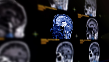
Intracranial monitoring is a test for people with epilepsy whose seizures cannot be controlled with medication. Intracranial monitoring helps doctors pinpoint where seizures are starting in the brain. In addition, the tests help “map” the brain, identifying areas that govern the brain’s essential functions. Neurosurgeons use these data in planning a patient’s epilepsy surgery.
What You Need to Know
- For intracranial monitoring, the surgeon places electrodes inside the skull to record electrical activity from the brain.
- Intracranial monitoring may be used to clarify a surgery plan when scalp electroencephalogram (EEG) findings are inconclusive or if different studies show conflicting results.
- Using data from intracranial monitoring helps brain surgeons more precisely locate and remove brain tissue that is causing seizures, while preserving essential brain functions such as speech and movement.
Why might my doctor recommend intracranial monitoring?
For some people with epilepsy, seizures are not resolved through medicine alone, and they may benefit from epilepsy surgery .
In diagnosing epilepsy and planning for surgery, a doctor is likely to order a range of tests to locate the cause of the seizures. Common noninvasive tests include an electroencephalogram (EEG) , magnetic resonance imaging (MRI) , positron emission tomography (PET) or single photon emission computed tomography (SPECT).
If these test results are inconclusive or conflicting, the doctor may recommend intracranial monitoring for a more detailed look at electrical activity in the brain. This involves surgically implanting electrodes inside the skull.
What are the risks of intracranial monitoring?
-
Bleeding
-
Infection
-
Brain edema (swelling)
-
Neurological impairments (rare)
What happens during intracranial monitoring for epilepsy?
Before the Procedure
Intracranial monitoring for epilepsy involves surgical implantation of electrodes inside the skull with the help of imaging guidance.
The surgical team members, including epilepsy specialists and neurosurgeons, neuropsychologists, neuroradiologists and others, review data from non-invasive tests and work together to plan the procedure.
A neurologist will take a careful history and examine you. If you are taking anti-seizure medications, the neurologist will help regulate them in preparation for the procedure. It is very important to tell your team about all the medicines you take, particularly any blood thinners including aspirin.
Implantation of Intracranial Electrodes
Once you are asleep under general anesthesia, the surgical team prepares the scalp and makes an incision in the skin. Depending on the type of electrodes being implanted, the surgical team will create one or more openings in the skull.
For stereo EEG or depth electrodes, the surgeon may drill a series of small burr holes. For electrode grids, the team may need to create one large surgical opening in the skull ( craniotomy ) so the grids can be placed across the surface of the brain.
The team uses imaging guidance to place the devices in the proper area beneath the skull, then secures them to surrounding tissue with sutures. The team then closes the surgical opening(s). A drain may be put in place, which is removed a day or two after the procedure.
Wires connecting the electrodes to external recording equipment are tunneled through the scalp and emerge through an incision in the skin. The team identifies each wire corresponding to an electrode so the recordings from those areas are accurate. The wires connect to a small portable pack.
After a short stay in the recovery room, you will go to the intensive care unit (ICU) or neurocritical care unit (NCCU) to spend the night.
Then, you will move to the epilepsy monitoring unit (EMU) , where the electrodes will be connected to equipment that continuously records your brain activity 24/7. You are free to walk around your room and use the bathroom; the portable pack that consolidates the electrode wires connects by a long cord to recording equipment that tracks your brain’s electrical signals as you move about.
The recording time typically lasts between three days and two weeks. The doctor may prescribe antibiotics during the recording period to lessen the risk of infection.
In the case of stereo EEG electrodes, the doctor removes these in the operating room when the monitoring is complete, and you return later for the surgery to address the cause of your epilepsy.