Ebstein Anomaly
What You Need to Know
- The age of onset for symptoms of Ebstein anomaly depends on the severity of the tricuspid valve defect.
- Newborn babies with severe persistent symptoms require surgery. Older infants and children may require tricuspid valve repair, depending on their symptoms.
- Long-term monitoring and follow-up are important in people with Ebstein anomaly, even in those who do not have any symptoms.
What is Ebstein anomaly?
Ebstein anomaly is a congenital (present at birth) heart defect of the tricuspid valve. The tricuspid valve is the inflow valve of the right lower chamber of the heart, which is called the right ventricle. Ebstein anomaly occurs when the tricuspid valve leaflets are formed abnormally; they are displaced downward into the right ventricle and stuck to the right ventricular wall. This results in “regurgitation” (leakage) of the tricuspid valve, which causes enlargement of the right upper chamber, called the right atrium, and decreased size of the right ventricle.
Children with Ebstein anomaly may also have additional heart defects, most commonly:
- Atrial septal defect: A hole between the two upper chambers of the heart
- Ventricular septal defect: A hole between the two lower chambers of the heart
- Pulmonary valve stenosis: The pulmonary valve (valve between the heart and blood vessel) to the lungs is thick and narrow, obstructing blood flow to the lungs.
- Pulmonary atresia: The pulmonary valve is closed off, preventing blood flow to the lungs from the heart. In this case, blood flow to the lungs is often through another blood vessel, the patent ductus arteriosus.
- Patent ductus arteriosus: An opening that causes blood to flow directly from the aorta (main artery of the body) to the pulmonary artery (main artery of the lungs).
What causes Ebstein anomaly?
The cause of Ebstein anomaly is not yet known. There is speculation that likely causes include genetic and environmental factors, such as exposure to lithium during pregnancy, though Ebstein anomaly can also occur in babies born to mothers who do not take lithium.
What are the symptoms of Ebstein anomaly?
The severity of the tricuspid valve defect will determine the types of symptoms and the age that those symptoms develop.
In the fetus:
- Severe tricuspid leakage may occur, preventing the right ventricle from pumping the blood forward.
- Hydrops, the accumulation of fluid in two or more compartments of the fetus’ body, may lead to swelling and possible heart failure.In severe cases, Ebstein anomaly may cause fetal death.
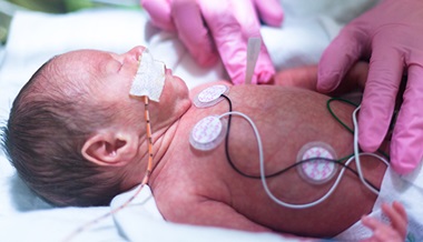
Newborn babies with severe tricuspid leaflet displacement may have cyanosis, a blue discoloration of the skin and lips due to low oxygen, after birth.
Children may first display symptoms, such as:
- shortness of breath
- cyanosis
- arrhythmias
- palpitations or fast heart rates
- In mild cases, Ebstein anomaly may be diagnosed when a heart murmur is heard during a routine physical examination.
How is Ebstein anomaly diagnosed?
There are several tests a pediatric cardiologist may use to diagnosis Ebstein anomaly. These can include:
- Obstetrical Ultrasound: Before birth, Ebstein anomaly may be detected during routine obstetrical ultrasound testing.
- Fetal Echocardiogram: Also used before birth, this provides doctors with a detailed ultrasound of the fetal heart.
- Transthoracic Echocardiogram: An ultrasound of the heart done after birth that demonstrates the anatomy and flow in the heart and blood vessels
- Electrocardiogram (ECG or EKG): A recording of the electrical conduction in the heart
- CT scan and MRI: 2D and 3D imaging of the heart anatomy, function and blood flow
How is Ebstein anomaly treated?
Management of Epstein Anomaly
Management of Ebstein anomaly will depend on age and severity of symptoms.
Before birth: Ongoing fetal echocardiograms will monitor the growth and well-being of the fetus and allow for careful planning of the delivery and any immediate care your baby will need after birth.
Newborns: When Ebstein anomaly is diagnosed after birth because a newborn displays cyanosis (blue discoloration due to low oxygen), the child will typically be admitted into an intensive care unit. Some newborns will have gradual improvement of cyanosis, while some may have persistent cyanosis or symptoms of heart failure and will require surgery before discharge.
Symptomatic babies and children may be initially managed with medications but will later need surgery to repair the tricuspid valve. Babies and children with arrhythmias may require medications and other treatments.
Surgery for Ebstein Anomaly
The timing and type of surgery depends on the severity of the tricuspid valve abnormality as well as the presence of additional heart defects.
Newborn babies with severe symptoms: These patients may require a systemic (body) to pulmonary (lungs) artery shunt. This is a surgically placed tube connecting a systemic blood vessel to the pulmonary artery to supply blood flow to the lungs. Babies born with the most severe defects may require a Starnes procedure, in which a patch is placed across the tricuspid valve in addition to placement of a systemic-to-pulmonary artery shunt.
Infants and older children with symptoms: Surgical repair of the tricuspid valve is usually done with the Cone Procedure, in which the surgeon detaches and reshapes the tricuspid valve leaflets to more closely align with the natural shape of the valve.
Older children: Surgical repair of the tricuspid valve may be recommended when there is progressive heart enlargement, exercise intolerance or cyanosis with exercise.
Children who have a mild tricuspid valve defect with a large atrial septal defect may benefit from closure of the defect. This can be done with a surgery that patches the hole or by cardiac catheterization where a device is guided into the heart through a catheter (a long, thin tube) and placed across the atrial septal defect to close the hole.
Children who have a severely abnormal tricuspid valve or who have not had good results from a tricuspid valve repair may require replacement of the tricuspid valve with a surgically placed artificial valve.