Retina
Our experts in the Retina Division treat retina-related disorders and conditions, such as macular degeneration, diabetic retinopathy, retinal detachment, vein occlusion and macular edema.
Learn more about the conditions we treat Discover our imaging
services
The Wilmer Difference More About Us
-
Our Experts
Meet the renowned team of retina experts who will help find the treatment that is best for you.
-
Our Research
Explore the groundbreaking research that is transforming the treatment of retinal diseases.
Conditions We Treat
The Retina Division provides expert clinical care of retina-related eye diseases and conditions including:
- Age related macular degeneration (AMD)
- Branch vein occlusion
- Central vein occlusion
- Choroidal melanoma
- Choroidal metastasis
- Choroidal neovascularization
- Choroidal hemangioma
- Choroidal nevus
- Diabetic retinopathy
- Epiretinal membrane
- Eye tumors
- Histoplasmosis
- Macular edema
- Macular hole
- Macular pucker
- Retinal detachment
- Retinitis pigmentosa
- Sickle cell retinopathy
- Trauma
- Vein occlusion
- Vitreous hemorrhage
Imaging Services Ophthalmic Ultrasound Service
The Ophthalmic Ultrasound Service at the Wilmer Eye Institute provides specialized diagnostic testing for diagnosing complex cases such as vitreous hemorrhage, retinal detachment, ocular tumors, optic disc edema and thyroid eye disease.
Conditions seen include:
- Vitreous Hemorrhage
- Endophthalmitis
- Intraocular and Orbital Tumors
- Iris and Ciliary Body Cysts and Lesions
- Dislocated Intraocular Lens
- Optic Disc Drusen and Papilledema
- Posterior Vitreous Detachment
- Retinal Detachment
- Choroidal Detachment
- Thyroid Eye Disease
- Posterior Scleritis
Services provided:
- Diagnostic B-Scan
- Diagnostic B-Scan with Qualitative A-Scan
- Ultrasound Biomicroscopy (UMB)
- Ultrasound Biometry
- Optical Biometry
Ophthalmic Photography Service
The Ophthalmic Photography Service at the Wilmer Eye Institute provides clinical and research related diagnostic images that are used by ophthalmic personnel in managing patients with various ocular conditions. The service offers multiple camera and imaging systems used to capture data. Ophthalmic photographers perform testing in clinics and operating rooms, work independently at Wilmer satellite locations, and participate in related investigative studies requiring protocol specific imaging.
Conditions seen include:
- Keratitis
- Cataracts
- Corneal Ulcer
- Diabetic Macular Edema
- Macular Degeneration
- Sickle Cell Retinopathy
Services provided:
- Color Retinal Fundus Photography
- Retinal Fluorescein Angiography
- ICG Angiography
- Slit Lamp Photography
- Intravenous Injection of Sodium Fluorescein And Indocyanine Green Dyes
- Ocular Coherence Tomography (OCT)
- Ocular Coherence Tomography Angiography (OCT-A)
- Corneal Specular Microscopy
Visual Physiology Service
The Visual Physiology Service at the Wilmer Eye Institute performs special tests to diagnose night blindness, color vision disorders, hereditary retinal disorders and other diseases. Often, these diseases cannot be diagnosed during routine examinations or require additional studies for the ophthalmologist to determine the correct diagnosis.
Conditions seen include:
- Stargardt disease
- Retinitis pigmentosa
- Macular dystrophies
- Retinal dystrophies
- Macular degeneration
- Age-related macular degeneration
Services provided:
- Comprehensive consultation and evaluation
- Electroretinograms (ERG, multifocal ERG)
- Electro-oculograms (EOG)
- Visual Evoked Responses (VEP)
- Color vision testing--D15, FM 100 Hue, Dark adapted perimetry
- Scanning laser ophthalmoscope macular perimetry and imaging
- Contrast Sensitivity
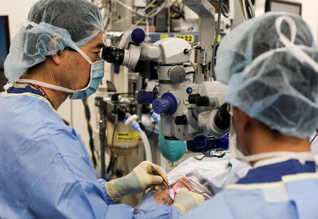
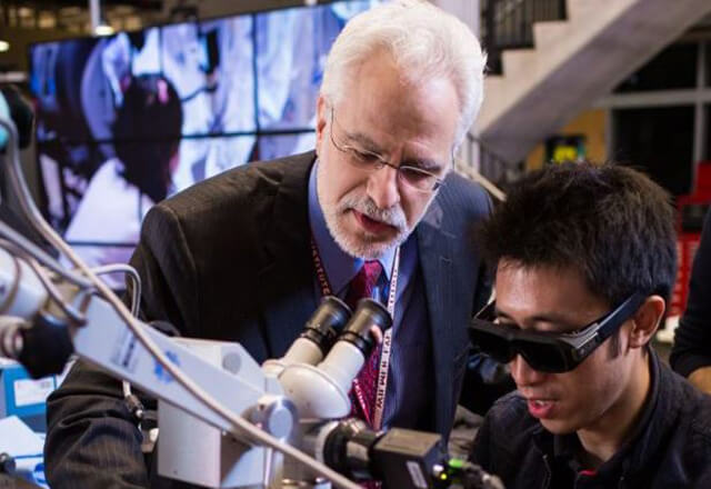
Our Innovative Treatments: Bionic Eye
Adolf Levi, 83, underwent the first bionic eye implant at Wilmer to restore his vision. Hear James Handa, M.D., discuss this extraordinary case.

Patient Stories
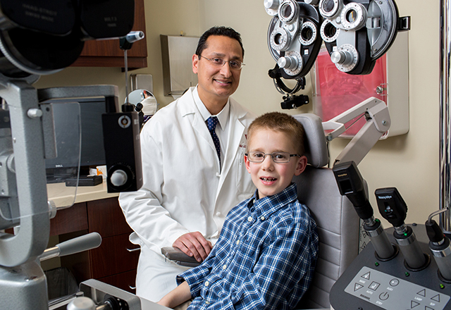
Coats' Disease: Sean's Story
At 5 years old, Sean was diagnosed with Coats' disease and went blind in one eye. Read the remarkable story of his sight restoration and his family's current commitment to supporting Coats' disease research.
Patient Perspective: Retinal Detachment
A detached retina can be frightening, but it is treatable. J. Myers shares his own experience, and his doctor, Wilmer Eye Institute retina specialist Dr. Adrienne Scott, provides insight into what occurs during retinal detachment and surgery to repair it. Read this patient's perspective.