-
Jeff Mumm, PhD

- Helen Larson & Charles Glenn Grover Professor in Ophthalmology
- Professor of Ophthalmology
Research Publications
-
Yanni Paulus, MD
- Chair, Continuing Medical Education Committee, Association for Research in Vision and Ophthalmology
- Associate Professor of Ophthalmology
Research Publications
-
Thomas Vincent Johnson, MD PhD

- Shelley and Allan Holt Rising Professor of Ophthalmology
- Associate Professor of Ophthalmology
Research Publications
Imaging is a central part of ophthalmology that include the reconstruction and analysis of spatial and temporal information in the eye over scales from molecules, subcellular components, cells to organs and whole populations. Imaging technology is highly interdisciplinary in nature, crossing physics, engineering, biology, pathology, and mathematics, and has several key components including:
- Imaging Technology: Optical, MRI, ultrasound, and molecular imaging
- Image Analysis: Image registration and reconstruction; extraction of knowledge from image data; machine learning and artificial intelligence
- Clinical and preclinical applications: Broad range of diagnostic and therapeutic applications enabled by advanced imaging technology
Research in Imaging technology
Our faculty are pioneering new imaging technologies, and novel applications in ophthalmology to advance our fundamental understanding of vision and clinical cares for ophthalmic conditions. Key research areas include:
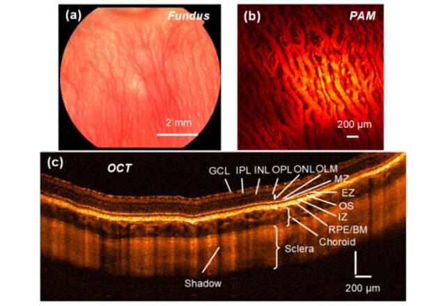
- Advanced Biophotonics: We are using novel optical imaging technologies, including visible light optical coherence tomography, photoacoustic microscopy, adaptive optics, two photo microscopy
- Image Analysis and Segmentation: We are applying mathematical models and machine learning to analyze information to understand disease and therapy responses, including using machine learning for imaging biomarker development.
- Imaging Algorithms :We are developing a range of physics or machine learning methods in computational imaging and advanced image reconstruction.
- Novel Imaging Systems: We are building new imaging technologies in a range of modalities.
Imaging Researchers


Imaging Research Labs at the Wilmer Eye Institute
-
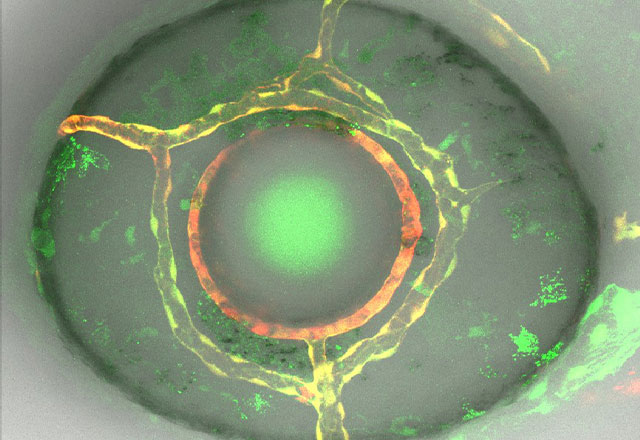
Mumm Lab
Dr. Mumm pioneered the use of multicolor microscopic imaging in the living retina to investigate how retinal neural circuits form during development. We now use this advanced imaging approach to visualize and quantify dynamic cellular interactions in the living retina.
-
Paulus Lab
The Paulus Advanced Retinal Imaging and Laser Laboratory is a dynamic, diverse, multidisciplinary group dedicated to improving the vision of patients suffering from eye diseases through applying biomedical engineering, lasers, photonics, optics, ultrasound, physics, nanoparticles, biochemistry, and mathematical modeling to develop novel retinal imaging systems and laser therapies.
-
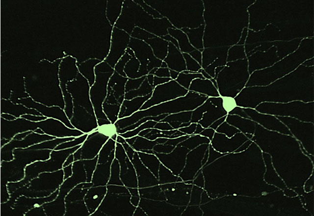
Johnson Lab
The Johnson Lab studies retinal ganglion cell transplantation as a potential therapeutic approach to restoring vision in glaucoma and other optic neuropathies.
Our Research in the Media
Life in action: Researchers capture 3D cellular dynamics across whole organism.PHYS.ORG (Winter 2022)
Oblique plane microscopy captures cell dynamics of whole organisms. BioTechniques (Winter 2022)
Smart Phone AI May Help Detect Diabetic Retinopathy. Netmeds.com (Spring 2021)