Research Lab Results
-
Daniel Nyhan Lab
The Daniel Nyhan Lab studies vascular changes that accompany aging to determine the underlying causes and find ways to reverse the process. One goal of our research is to identify the factors that cause vascular stiffness. Our hope is that our work in vascular biology will lead to new ways to improve vascular compliance and thereby improve cardiovascular function and perioperative risk.
-
Wei Dong Gao Lab
Work in the Wei Dong Gao Lab primarily focuses on heart failure and defining molecular and cellular mechanisms of contractile dysfunction. We use molecular biology and proteomic techniques to investigate the changes that myofilament proteins undergo during heart failure and under drug therapy. We're working to determine the molecular nature of nitroxyl (HNO) modification of tropomyosin. -
Nicola Heller Lab
Research in the Nicola Heller Lab focuses on the immunobiology of macrophages. Our team explores how these cells impact diseases with an inflammatory element, such as cancer, cardiovascular disease and obesity. Using a variety of techniques, including molecular and cellular biology, biochemistry, mouse models and more, we study the role of IL-4/IL-13 signaling in asthma and allergic disease, as well as the role of alternatively activated macrophages (AAM) in the pathogenesis of allergic inflammation. Currently, we are researching the links between asthma and obesity, with a focus on the roles of gender and race. -
Nathaniel Comfort Lab
Research in the Nathaniel Comfort Lab looks at the history of biology. Areas of particular interest include heredity and health in 20th century America, genetics, molecular biology, biomedicine, the history of recent science, oral history and interviewing. -
O'Rourke Lab
The O’Rourke Lab uses an integrated approach to study the biophysics and physiology of cardiac cells in normal and diseased states. Research in our lab has incorporated mitochondrial energetics, Ca2+ dynamics, and electrophysiology to provide tools for studying how defective function of one component of the cell can lead to catastrophic effects on whole cell and whole organ function. By understanding the links between Ca2+, electrical excitability and energy production, we hope to understand the cellular basis of cardiac arrhythmias, ischemia-reperfusion injury, and sudden death. We use state-of-the-art techniques, including single-channel and whole-cell patch clamp, microfluorimetry, conventional and two-photon fluorescence imaging, and molecular biology to study the structure and function of single proteins to the intact muscle. Experimental results are compared with simulations of computational models in order to understand the findings in the context of the system as a whole. Ongoing studies in our lab are focused on identifying the specific molecular targets modified by oxidative or ischemic stress and how they affect mitochondrial and whole heart function. The motivation for all of the work is to understand • how the molecular details of the heart cell work together to maintain function and • how the synchronization of the parts can go wrong Rational strategies can then be devised to correct dysfunction during the progression of disease through a comprehensive understanding of basic mechanisms. Brian O’Rourke, PhD, is a professor in the Division of Cardiology and Vice Chair of Basic and Translational Research, Department of Medicine, at the Johns Hopkins University.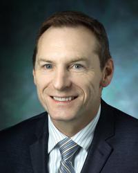
-
Fu Lab
The Fu Lab is a basic research lab that studies zinc transport, with a particular focus on which step in the zinc transport process may be modulated and how. Dr. Fu's lab uses parallel cell biology and proteomic approaches to understand how these physiochemical principles are applied to mammalian zinc transporters and integrated to the physiology of pancreatic beta cells. This research has implications for understanding how zinc transport is related to diabetes and insulin intake. -
Inoue Lab
Complexity in signaling networks is often derived from co-opting one set of molecules for multiple operations. Understanding how cells achieve such sophisticated processing using a finite set of molecules within a confined space--what we call the ""signaling paradox""--is critical to biology and engineering as well as the emerging field of synthetic biology. In the Inoue Lab, we have recently developed a series of chemical-molecular tools that allow for inducible, quick-onset and specific perturbation of various signaling molecules. Using this novel technique in conjunction with fluorescence imaging, microfabricated devices, quantitative analysis and computational modeling, we are dissecting intricate signaling networks. In particular, we investigate positive-feedback mechanisms underlying the initiation of neutrophil chemotaxis (known as symmetry breaking), as well as spatio-temporally compartmentalized signaling of Ras and membrane lipids such as phosphoinositides. In parallel, we also try to understand how cell morphology affects biochemical pathways inside cells. Ultimately, we will generate completely orthogonal machinery in cells to achieve existing, as well as novel, cellular functions. Our synthetic, multidisciplinary approach will elucidate the signaling paradox created by nature. -
Mahendra Damarla Lab
Work in the Mahendra Damarla Lab focuses primarily on the field of vascular biology. Much of our research involves exploring alternatives to mechanical ventilation as a therapy for acute lung injury. We investigate mitogen-activated protein kinase-activated protein kinase 2 as a method to mediate apoptosis during lung vascular permeability by regulating movement of cleaved caspase 3. We have also conducted research on the prevalence of confirmatory tests in patients hospitalized with congestive heart failure or chronic obstructive pulmonary disease (COPD).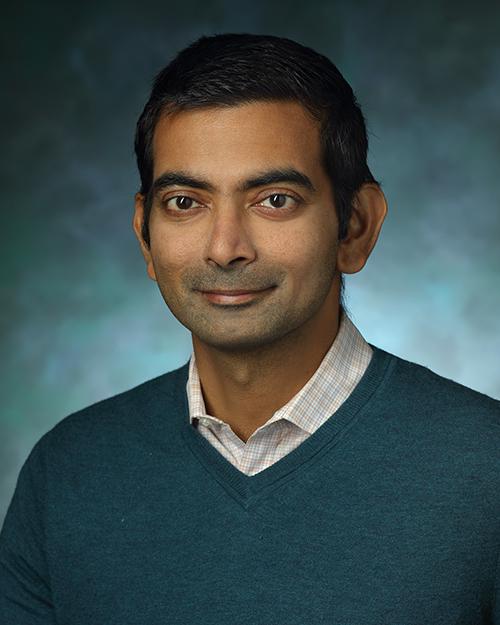
-
The Pathak Lab
The Pathak lab is within the Division of Cancer Imaging Research in the Department of Radiology and Radiological Science. We develop novel imaging methods, computational models and visualization tools to ‘make visible’ critical aspects of cancer, stroke and neurobiology. Our research broadly encompasses the following areas: Functional and Molecular Imaging; Clinical Biomarker Development; Image-based Systems Biology and Visualization and Computational Tools. We are dedicated to mentoring the next generation of imagers, biomedical engineers and visualizers. Additional information can be found at www.pathaklab.org or by emailing Dr. Pathak. -
Shanthini Sockanathan Laboratory
The Shanthini Sockanathan Laboratory uses the developing spinal cord as our major paradigm to define the mechanisms that maintain an undifferentiated progenitor state and the molecular pathways that trigger their differentiation into neurons and glia. The major focus of the lab is the study of a new family of six-transmembrane proteins (6-TM GDEs) that play key roles in regulating neuronal and glial differentiation in the spinal cord. We recently discovered that the 6-TM GDEs release GPI-anchored proteins from the cell surface through cleavage of the GPI-anchor. This discovery identifies 6-TM GDEs as the first vertebrate membrane bound GPI-cleaving enzymes that work at the cell surface to regulate GPI-anchored protein function. Current work in the lab involves defining how the 6-TM GDEs regulate cellular signaling events that control neuronal and glial differentiation and function, with a major focus on how GDE dysfunction relates to the onset and progression of disease. To solve these questions, we use an integrated approach that includes in vivo models, imaging, molecular biology, biochemistry, developmental biology, genetics and behavior.