Research Lab Results
-
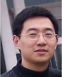
Jun Hua Lab
Dr. Hua's research has centered on the development of novel MRI technologies for in vivo functional and physiological imaging in the brain, and the application of such methods for studies in healthy and diseased brains. These include the development of human and animal MRI methods to measure functional brain activities, cerebral perfusion and oxygen metabolism at high (3 Tesla) and ultra-high (7 Tesla and above) magnetic fields. He is particularly interested in novel MRI approaches to image small blood and lymphatic vessels in the brain. Collaborating with clinical investigators, these techniques have been applied 1) to detect functional, vascular and metabolic abnormalities in the brain in neurodegenerative diseases such as Huntingdon's disease (HD), Parkinson's disease (PD), Alzheimer's disease (AD) and mental disorders such as schizophrenia; and 2) to map brain functions and cerebrovascular reactivity for presurgical planning in patients with vascular malformations, brain tumors and epilepsy. -
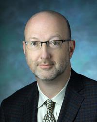
James Pekar Lab
How do we see, hear, and think? More specifically, how can we study living people to understand how the brain sees, hears, and thinks? Recently, magnetic resonance imaging (MRI), a powerful anatomical imaging technique widely used for clinical diagnosis, was further developed into a tool for probing brain function. By sensitizing magnetic resonance images to the changes in blood oxygenation that occur when regions of the brain are highly active, we can make ""movies"" that reveal the brain at work. Dr. Pekar works on the development and application of this MRI technology. Dr. Pekar is a biophysicist who uses a variety of magnetic resonance techniques to study brain physiology and function. Dr. Pekar serves as Manager of the F.M. Kirby Research Center for Functional Brain Imaging, a research resource where imaging scientists and neuroscientists collaborate to study brain function using unique state-of-the-art techniques in a safe comfortable environment, to further develop such techniques, and to provide training and education. Dr. Pekar works with center staff to serve the center's users and to keep the center on the leading edge of technology. -
Janet Record Lab
Research in the Janet Record Lab focuses on medical education and patient-centered care. We’re currently developing a curriculum for internal medicine residents in the inpatient general medicine service setting. The curriculum teaches residents to use hand-carried ultrasound for imaging the inferior vena cava to assess volume status.
-
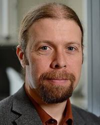
Jinyuan Zhou Lab
Dr. Zhou's research focuses on developing new in vivo MRI and MRS methodologies to study brain function and disease. His most recent work includes absolute quantification of cerebral blood flow, quantification of functional MRI, high-resolution diffusion tensor imaging (DTI), magnetization transfer mechanism, development of chemical exchange saturation transfer (CEST) technology, brain pH MR imaging, and tissue protein MR imaging. Notably, Dr. Zhou and his colleagues invented the amide proton transfer (APT) approach for brain pH imaging and tumor protein imaging. His initial paper on brain pH imaging was published in Nature Medicine in 2003 and his most recent paper on tumor treatment effects was published in Nature Medicine in 2011. A major part of his current research is the pre-clinical and clinical imaging of brain tumors, strokes, and other neurologic disorders using the APT and other novel MRI techniques. The overall goal is to achieve the MRI contrast at the protein and peptide level without injection of exogenous agents and improve the diagnostic capability of MRI and the patient outcomes.
-
J. Webster Stayman Lab
The J. Webster Stayman Lab studies both emission tomography and transmission tomography (CT, tomosynthesis and cone-beam CT). Our research activities relate to 3-D reconstruction, including model-based statistical / iterative reconstruction, regularization methods and modeling of imaging systems. We are developing a generalized framework for penalized likelihood (PL) reconstruction combining statistical models of noise and image formation with incorporation of prior information, including patient-specific prior images, atlases and models of components / devices known to be in the field of view. Our research includes algorithm development and physical experimentation for imaging system design and optimization. -
CORE-320 Multicenter Trial Lab
The central theme of the CORE-320 Multicenter Trial Lab’s research is to support the Coronary Artery Evaluation Using 320-Row Multidetector CT Angiography (CORE 320) study, a multi-center multinational diagnostic study with the primary objective to evaluate the diagnostic accuracy of 320-MDCT for detecting coronary artery luminal stenosis and corresponding myocardial perfusion deficits in patients with suspected CAD compared with the reference standard of conventional coronary angiography and SPECT myocardial perfusion imaging. Armin Arbab-Zadeh, MD, PhD, is an associate professor of medicine at the Johns Hopkins University School of Medicine and Director of Cardiac Computed Tomography in the Division of Cardiology at the Johns Hopkins Hospital in Baltimore. Research Areas: coronary/cardiac imaging, coronary risk prediction, heart attack prevention, cardiac computed tomography, coronary circulation and disease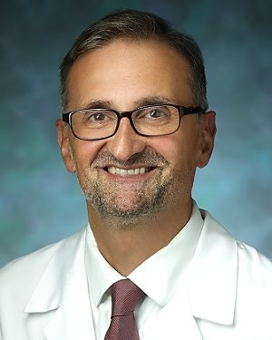
-
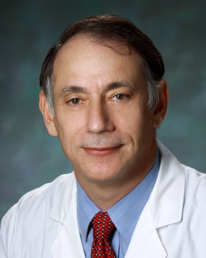
Cardiology Bioengineering Laboratory
The Cardiology Bioengineering Laboratory, located in the Johns Hopkins Hospital, focuses on the applications of advanced imaging techniques for arrhythmia management. The primary limitation of current fluoroscopy-guided techniques for ablation of cardiac arrhythmia is the inability to visualize soft tissues and 3-dimensional anatomic relationships. Implementation of alternative advanced modalities has the potential to improve complex ablation procedures by guiding catheter placement, visualizing abnormal scar tissue, reducing procedural time devoted to mapping, and eliminating patient and operator exposure to radiation. Active projects include • Physiological differences between isolated hearts in ventricular fibrillation and pulseless electrical activity • Successful ablation sites in ischemic ventricular tachycardia in a porcine model and the correlation to magnetic resonance imaging (MRI) • MRI-guided radiofrequency ablation of canine atrial fibrillation, and diagnosis and intervention for arrhythmias • Physiological and metabolic effects of interruptions in chest compressions during cardiopulmonary resuscitation Henry Halperin, MD, is co-director of the Johns Hopkins Imaging Institute of Excellence and a professor of medicine, radiology and biomedical engineering. Menekhem M. Zviman, PhD is the laboratory manager. -
Accessible Magnetic Resonance Laboratory
VISION: To make MRI more equitable and inclusive
MISSION: To develop and deploy MRI tools and methods to enable accessible imaging of underserved populations
-
Clifton O. Bingham III Lab
Research in the Clifton O. Bingham III Lab focuses on defining clinical and biochemical disease phenotypes related to therapeutic responses in rheumatoid arthritis and osteoarthritis; developing rational clinical trial designs to test new treatments; improving patient-reported outcome measures; evaluating novel imaging modalities for arthritis; and examining the role of oral health in inflammatory arthritis.
-
The Bigos Lab
The Bigos Lab focuses on a Precision Medicine approach to the treatment of psychiatric illness. In addition, this lab employs functional neuroimaging and genetics as biomarkers in neuropsychiatric drug development. A recent study used functional MRI to test the neural effects of a drug with the potential to treat cognitive dysfunction in schizophrenia. Other studies aim to identify patient-specific variables including sex, race, and genetics that impact drug clearance and clinical response to better select and dose antipsychotics and antidepressants.