COVID-19 Research Recap
During the COVID-19 pandemic, Johns Hopkins Radiology has continued its research efforts while prioritizing the health and safety of all researchers, staff, and community. Because of COVID-19 all but the most critical in-person research activities were shut down in March. This did not deter Radiology research faculty, fellows and staff who remained active throughout the shutdown period, analyzing data, publishing manuscripts, submitting grant applications and progress reports, giving virtual talks, and attending virtual meetings. Essential personnel came in to make sure that service center instruments and equipment were safely maintained, liquid nitrogen cell freezers were filled, and that the lab equipment was running smoothly in readiness for a return to work.
Radiology faculty members actively participated in the virtual ISMRM meeting that was held in August. Several scientific presentations and talks were given by the faculty.
Click on the links below to learn more about our research efforts during this time and how to contribute.
Learn about some of our work:
Research Funding
Below are the new grants awarded from April 2020 to June 2020 within the Johns Hopkins Radiology department.
| Title | Sponsor | PI |
|---|---|---|
|
Radiology Medical Education and Mentoring Grant |
M.R. and Evelyn Hudson Foundation |
Donna Magid |
|
Intra-Operative 4-D Soft Tissue Perfusionusing No Gantry Rotation (Ipen) |
National Institute of Biomedical Imaging and Bioengineering |
Katsuyuki Taguchi |
|
Ultrafast Quantitative Ph MRI For Acute Ischemic Stroke Patients |
National Inst Of Neurological Disorders |
Hye Young Heo |
|
In Vivo Evaluation of NM21-1601 Using PD-L1 PET |
Numab Therapeutics Ag |
Sridhar Nimmagadda |
|
Non-Invasive Imaging Studies of NVD003 |
Novadip Biosciences Sa |
Jeff Bulte |
|
Quasi-Ideal Photon Counting X-Ray CT with Multi-Energy Inter-Pixel Coincidence |
National Institute of Biomedical Imaging and Bioengineering |
Katsuyuki Taguchi |
|
Multi-Voxel Spectral Editing At 3T |
National Institute of Biomedical Imaging and Bioengineering |
Peter Barker |
|
Ultra Coils Form Start to Finish For The Endovascular Repair Of Small Intracran |
Univ Of Maryland Baltimore |
Ferdinand Hui |
|
Disseminating A Patient Centered Venous Thromboembolism Prevention Bundle |
Agency for Healthcare Research and Quality |
Brandyn Lau |
|
Optoacoustic Imaging Scanner |
Office of The Director Nih |
Zaver Bhujwalla |
|
MRI-Guided Cryoablation And Imaging-Guided Intratumoral Delivery Of STING |
National Cancer Institute |
Dara Kraitchman |
|
Non-Contrast MR Imaging of Blood-Brain-Barrier Permeability In Alzheimer's Disea |
National Institute on Aging |
Hanzhang Lu |
|
The Tumor Microenvironment in Nanoparticle Delivery And Function |
National Cancer Institute |
Zaver Bhujwalla |
|
Development Of 5D3 Mab and USPIO-Based Theranostics For Image-Guided Prostate... |
Congressionally Directed Medical Research Programs |
Sudath Hapuarachchige |
COVID-19 Research Working Group

The immediate consequences of COVID-19 have been clearly visible in recent months with lists of common symptoms and first-hand accounts circulating. It is the long-term and chronic consequences of this new virus that remains unknown and will need further research.
Appropriately, new funding avenues have opened up, including internal funding from Johns Hopkins University. Within the department of radiology, several faculty members have separately submitted applications which led to the realization that future coordination of efforts within and outside the department would assist in sharing new information.
Dr. Hanzhang Lu is currently heading the Radiology COVID-19 Research Working Group which aims to provide a visible platform for information sharing and quick access to the latest developments in this field. This group would not only help create a launching pad for future research applications based on NIH news and announcements but also provide regular virtual meetings with speakers inside and outside the department to discuss imaging’s role in future projects. The first meeting was held on July 8 with a presentation given by an infectious disease specialist at Johns Hopkins. Future meetings are being planned.
An initial email inviting faculty members resulted in an enthusiastic response with 28 starting members from across all divisions and modalities. These members will begin gathering preliminary data and formulating hypotheses and projects for anticipated grant applications. COVID-19 is a novel disease and Dr. Lu states that “we are all learning together so we can better communicate ideas and share knowledge to foster collaborations and techniques.” Any faculty members interested in joining are welcome to contact Dr. Lu at [email protected].
Anticipated Applications for MPI Scanner
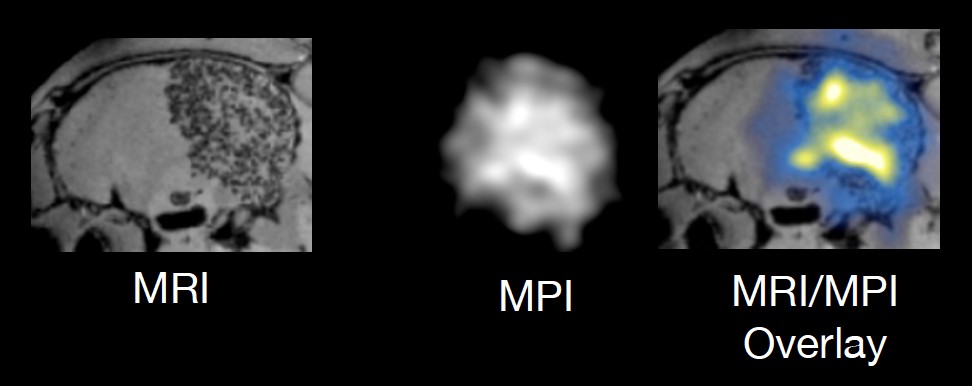
Unilateral homing of magnetic nanoparticles can be seen on the MRI as hypointense spots and on the corresponding MPI as hot spots without background signal. (Unpublished) data were obtained with a Momentum beta-version prototype MPI scanner.
The COVID-19 pandemic had an immediate impact on research progress as all but the most critical research activity was restricted. One of the many delays included the installation of the MPI scanner at Kennedy Krieger Institute which has been pushed to late fall/early winter. This MPI scanner, the first on the East Coast, was acquired through a successful NIH S10 shared instrumentation grant awarded to Dr. Jeff Bulte in 2019. Now, this revolutionary imaging modality can be applied for a vast array of studies including cell tracking and inflammation imaging which offers COVID-19 related applications.
The MPI scanner offers major applications through its unique sensitivity for magnetic nanoparticles. The first is tracking and labeling of therapeutic cells to target tumors or repair tissue and thereby see the progress of cells within the body. It can also monitor particles as they circulate in blood flow for functional brain imaging and vascular profusion and malformation imaging. Another application is through magnetic hyperthermia where magnetic particles are heated with an electric field for precision-based tumor targeting. This MPI has an add-on module for magnetic heating to monitor the particles and tumor progress.
The imaging of inflammation by injecting particles to track macrophages, the first cells at areas of inflammation, offers key visualization for many chronic diseases including multiple sclerosis, Alzheimer’s, and potentially COVID-19.
As construction slowly resumes, Dr. Bulte continues to procure materials and test particles to see the signals they can give for the MPI scanner to pick up in future studies. He states that “COVID-19 will continue to be very important and we must keep up our established research program to continue work on the many chronic conditions that can benefit from molecular imaging.”
Projects on the MPI scanner are partnered with 11 investigators across Johns Hopkins and Kennedy Rieger Institute for interdisciplinary, precision-based research that includes PIs from biomedical engineering and radiation oncology departments.
Optoacoustic Scanner
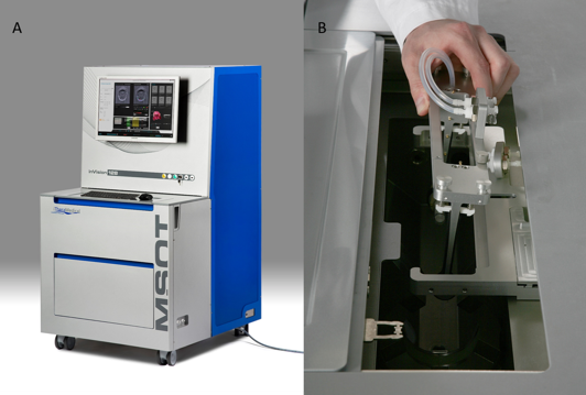
Representative picture of (A) the inVision 512-echo MSOT system and (B) the animal holder that is positioned in the imaging chamber for in vivo MSOT imaging.
In an multidisciplinary team effort under the leadership of Dr. Bhujwalla, investigators from Radiology and six other departments across JHU (Drs. Artemov, Bell, Bhujwalla, Bulte, Casero, Ding, Gabrielson, Gerecht, Hanes, Liu, Nimmagadda, O’Rourke, Pomper, Tran and Wang) successfully submitted an NIH High-End Instrument application, to obtain funding to purchase the scanner. The scanner is slated to be installed in September 2020 in dedicated space in our brand new satellite facility in Cancer Research Building 2 (CRB2). The facility has a built-in animal holding area to house experimental animals. The animal vivarium is located directly above, allowing for easy transfer of animals to the imaging center. An advanced small animal radiation research platform (SARRP) is located adjacent to this satellite facility. Given the ability of OA imaging to detect oxygenation and hemodynamics, the location of the SARRP is strategically useful as it will significantly advance radiation therapy research.
The MSOT scanner will significantly advance the ability of JHU Faculty to understand and treat human diseases. Integrating the capabilities of the MSOT scanner within the multimodal imaging abilities of the MRB Molecular Imaging Service Center and Cancer Functional Imaging Core will significantly advance current and future research efforts of the faculty. The ability, in the future, to attach a handheld device to the MSOT scanner, creates the possibility of translating the research being conducted to large animals, and to humans, once iThera obtains FDA approval for the handheld device.
The iThera MSOT inVision 512-echo scanner provides versatile specificity of optical contrast with high spatiotemporal real-time resolution imaging of optical absorbers, of both intrinsic (e.g. hemoglobin) and extrinsically administered (e.g. indocyanine green) chromophores. In addition, this instrument allows visualization of tissue structures using conventional ultrasound imaging. This is the first system of its kind that simultaneously delivers functional, anatomical, and molecular imaging in real time within a whole body small animal imaging system. The MSOT inVision 512-echo is the first and only tomographic OA imaging system that integrates simultaneous tomographic optoacoustic and tomographic reflection mode ultrasound acquisitions. The ultrasound imaging capacities exceed performance of the common linear-array-based probes by delivering entire cross-sectional views of the animal without the need for physical contact (and deformation) with the animal. Due to the lack of contact and the fixed scanning geometry, the quality and utility of the acquired ultrasound images do not depend on the skill of the operator. The instrument is equipped with 360o laser Illumination of the animal and produces a homogenous light field for even distribution of light through tissue at the full depth within a small animal, a function necessary for whole-body imaging. The tunable laser allows generation of light from 680-980 nm as standard with options for extending wavelengths ranging from 660-1300 nm. The capacity for continuous imaging from 660-1300 nm is a unique feature allowing optimal imaging of red absorbers (e.g. iRFP), collagen (at 1030 nm), and lipids (at 1064 nm). Spectra of unknown substances can be unmixed from intrinsic signals (typically Hb, HbO2 and melanin) and stored for future use.