Melanoma Prevention & Diagnosis
This year, it is estimated that more than three million people will be diagnosed with skin cancer, and more than 70,000 people will be diagnosed with melanoma, the most deadly form of skin cancer. Johns Hopkins dermatologists provide extensive evaluations of the skin. While Johns Hopkins scientists are discovering new ways to diagnose, treat, and prevent skin cancers, there are several proactive steps that our experts suggest to help reduce the risk of getting this disease.
Sun Safety Tips
Sunscreen: Slather it on! Apply a broad-spectrum sunscreen that protects against UVA and UVB rays, with an SPF 30 or higher, liberally and frequently. Remember that sunless tanning products do not contain enough sunscreen to be protective.
Sunbathers Beware: Exposure to the sun for long periods can increase your risk of developing a skin cancer dramatically, especially during the sun’s peak hours of 10 am – 3 pm. So-called “base” tans do not offer protection from burns, nor do they extend the time-to-burn.
Stay Away From Tanning Beds and Sun Lamps: There is no safe type of UV radiation, despite manufacturer’s claims. These devices also can impair vision and age the skin prematurely.
Check/Examine Your Skin: Do you have a funny-looking mole or a history of skin cancer in your family? Melanoma can appear anywhere on the skin’s surface – even on normal-appearing skin. The Skin Cancer Foundation recommends that everyone practice a head-to-toe self-examination of their skin once a month to look for any new or changing areas. Most people can follow the ABCDEs of melanoma:
- A: Asymmetric
- B: Borders that are irregular
- C: Color that is unusual or varied
- D: Diameter more than 6mm, or the size of a pencil eraser
- E: Evolving or changing moles.
People with ethnic skin tones, these rules may not apply. People of color may develop melanoma in different spots, including the eyelids, nails, genitals adn in the mouth.
Other potential warning signs of skin cancer include:
- A skin growth that increases in size and appears pearly, translucent, tan, brown, black, or multicolored
- A spot or sore that continues to itch, hurt, crust, scab, erode, or bleed
- An open sore that does not heal within three weeks
If you see something that looks like skin cancer, or are concerned about any spots, make an appointment to see a dermatologist. The earlier skin cancer is diagnosed and treated, the more likely it can be cured. Learn more about skin cancer prevention and screening.
Diagnosis
Biopsies
Cutaneous melanoma can appear anywhere on the skin surface. Melanoma can develop in a pre-existing mole, or arise on normal-appearing skin. It is suspected when a “mole” looks uneven in terms of its border, shape, or color. Diagnosis is confirmed with a simple skin biopsy, which is performed using one of several techniques. The American Academy of Dermatology has more information on the clinical appearance of melanoma.
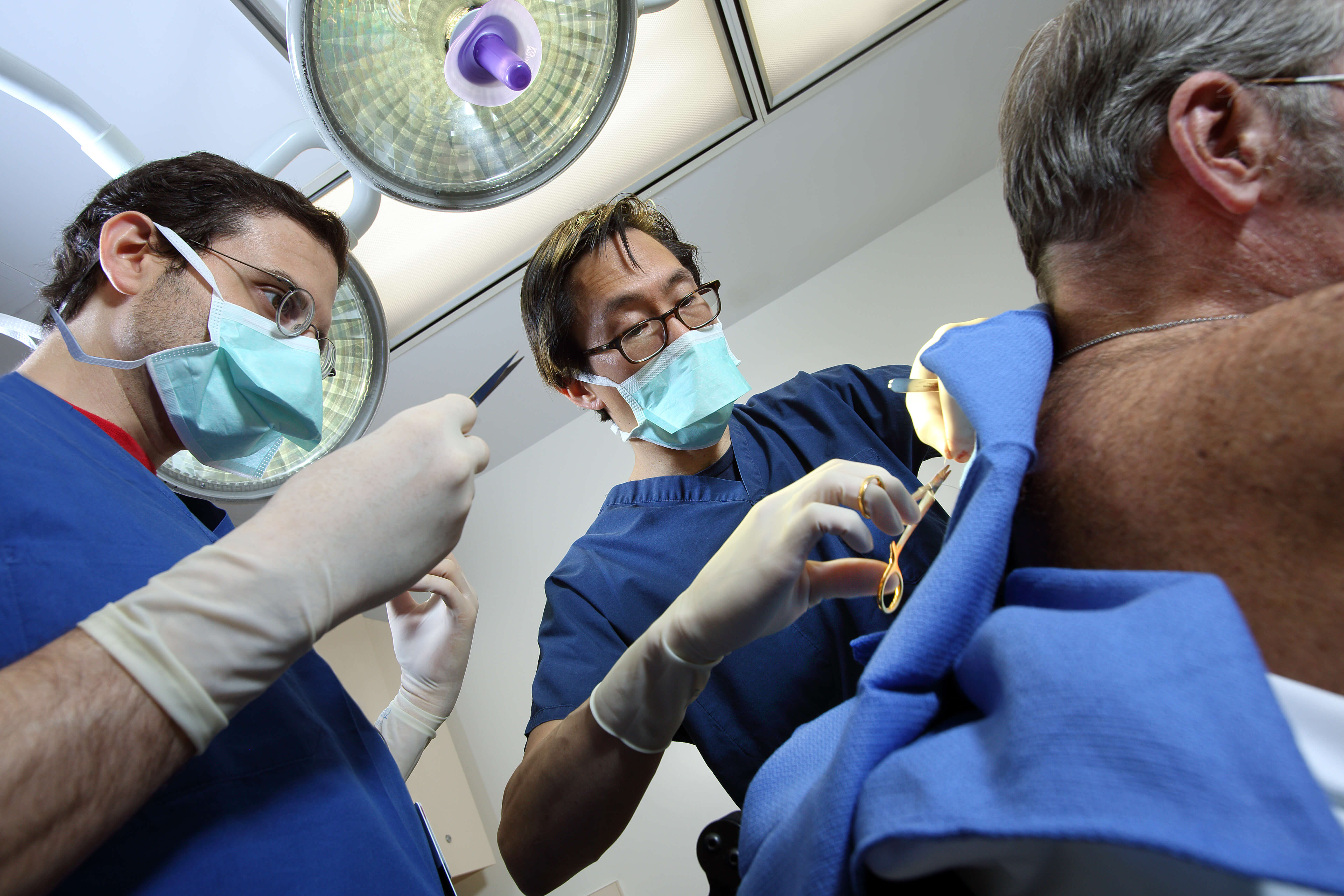
Excisional/Incisional Biopsy
An excisional biopsy is the removal of an entire skin lesion with a tiny rim of normal-looking skin. It is a common method for collecting a skin sample to determine whether it is cancerous. This technique leaves a small scar. An incisional biopsy removes a small part of the lesion for testing. This is often done using local anesthesia to numb the area so patients do not feel pain.
Special Sites
In rare cases, where a melanoma is suspected in other locations such as the nail bed, special techniques may be used to sample or remove the lesion for testing. In the case of oral or genital mucosal lesions, small biopsies should be performed just like skin lesions.
Fine-Needle Aspiration
This technique uses a thin needle and syringe to remove small amounts of fluid or tissue from suspected tumors. It is not used for a primary melanoma site, but for a discrete lump of mass that could represent melanoma recurrence or melanoma that has spread from the skin to a lymph node or other area. In some circumstances, an ultrasound or computed tomography-guided fine needle aspiration may be appropriate if the area is best seen on imaging studies.
Understanding Your Pathology Report
For primary melanoma, one of the most important factors is tumor thickness, which is a surrogate for tumor volume. Tumor depth is the strongest prognostic indicator. The next factors that are additive risk indicators include ulceration over most of the tumor surface, rate of mitosis (cell division), presence of satellite tumor spread and nerve invasion.
At Johns Hopkins, dermatopathologists physicians who specialize in diagnosing diseases of the skin, review all tissue and fluid samples to determine presence of melanoma, and send a report to the primary physician. Your doctor will then go over the results of your pathology report and answer any questions.