Because of the fact that vascular malformations are rare and because of the precision required to treat them, our interventional radiologists work within the Vascular Anomalies Center to offer patients individualized treatment plans.
Vascular Malformations: Why Choose Johns Hopkins
Our interventional radiologists Clifford Weiss and Brian Holly specialize in diagnosing and treating all forms of vascular malformations.
We also have a group of physicians who manage patients with PAVMs and Hereditary Hemorrhagic Telangiectasia (HHT), consisting of pulmonary specialists, ear nose and throat (ENT) specialists, neuro specialists, geneticists, and interventional radiology.
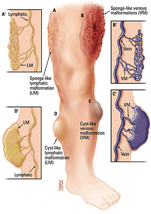
Vascular malformation is a general term that includes congenital vascular anomalies of only veins, only lymph vessels, both veins and lymph vessels, or both arteries and veins.
Both veins and lymph vessels: venolymphatic malformations (VLM)
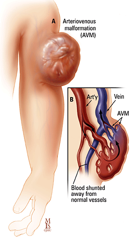
Arteries connected directly to veins without any capillaries in between: arteriovenous malformation (AVM)
Why do these vascular malformations occur?
These are all present at birth and become apparent at different ages. We are just beginning to understand how malformations occur. The pulmonary arteriovenous malformation, when associated with Hereditary Hemorrhagic Telangiectasia (HHT), is inherited genetically. There is currently much work being done on the possible genetics of other malformations. Most are only known as something that occurs during development of the arteries, veins, and/or lymph vessels, but without specific cause.
What are the symptoms of a vascular malformation?
These vascular malformations can cause a variety of symptoms depending on the location in the body:
Venous malformation may cause pain where ever they are located. Venous and lymphatic malformations may cause a lump under the skin. There may be an overlying birthmark on the skin. Bleeding or lymph fluid leaking may occur from skin lesions. Lymphatic malformations tend to become infected, requiring repeated antibiotic treatments. Venous and lymphatic malformations may be associated with a syndrome called Klippel-Trenaunay Syndrome.
Arteriovenous malformations may cause pain. They are also more stressful on the heart because of the rapid shunting of blood from arteries to veins. Depending on their location, they may also result in bleeding (for example from the bowels, from the uterus or from the bladder).
Hemangioma is another common term used for vascular anomalies. However, this name actually applies to a childhood vascular anomaly that has a rapid growth phase between birth and 3 months of age. These will resolve completely by age 7. The major reason for us to treat these is for low platelets that do not respond to medical treatment, or in the liver because of massive shunting with a strain on the heart.
Pulmonary arteriovenous malformations (PAVMs) are somewhat different in that they shunt blood from the right heart system to the left heart system without picking up oxygen in the lungs. This results in symptoms of low oxygen, shortness of breath, fatigue. These malformations may also bleed, resulting in coughing up blood or blood in the chest. Also, these pulmonary artery to pulmonary vein shunts can allow clots to pass through the lungs and travel to the arteries in the body, with risk for stroke or brain abscess. This is a very significant reason to block these shunts in anyone who has a non inherited pulmonary arteriovenous malformation (PAVM) or anyone who has inherited PAVMs with the syndrome of HHT [HHT has another name: OWR - Osler Weber Rendu].
Program Director, Interventional Radiology Integrated Residency
Assistant Professor of Radiology and Radiological Science
About Vascular Malformations and Your Treatment
These can often be seen on physical examination. Deeper vascular malformations can be diagnosed on MRI (magnetic resonance imaging).
Although surgery is sometimes useful, it is usually difficult for surgeons to completely remove vascular malformations, which will return if not removed completely. A nonsurgical method of closing down the blood or lymph flow into the malformation is done by interventional radiologists, who treat patients with image guided procedures. Vascular malformations are treated by embolization. The AVMs and hemangiomas can be closed by advancing a tiny plastic tubing, no larger than a pencil point, into the feeding artery to the malformation. This can be done without incisions or stitches, and with only mild sedation. Medical glue or alcohol or small beads are then floated into the malformation until it is full and no longer has blood flowing through it. For Pulmonary AVMs platinum coils are used to block flow through the feeding artery to the malformation. The VMs and LMs are closed by injecting alcohol into the sacs filled with venous blood or lymph until these sacs collapse and no longer fill.
The arteriovenous malformations can be treated with a one-night hospital stay. There is usually minimal discomfort for one to three days. The venous and lymphatic malformations also require one night in the hospital. These malformations swell after treatment with alcohol, and the swelling and pain may last for three to five days. During this time, we give patients medication for any pain or swelling they may have. The full shrinkage of these malformations may take four to six weeks.
Pulmonary arteriovenous malformations are very effectively treated by embolization which blocks only the abnormal artery feeder, and preserves the normal lung arteries. Patients usually notice an immediate improvement in symptoms with the immediate increase in oxygen level. Other arteriovenous malformations are more difficult to treat since they tend to pull in new artery feeders from time to time. However, embolization is very effective in blocking abnormal artery feeders while preserving normal arteries. AVMs may require a series of treatments to block all of the abnormal feeders. Venous and lymphatic malformations respond well to alcohol embolization. These may also require a series of treatments about six weeks apart to block all of the abnormal vessels. All vascular malformations require long term surveillance, so that if there is any change, such as a growth spurt with puberty or pregnancy or menopause, they can be monitored for symptoms that may warrant rechecking and possibly retreating. We monitor PAVMs over time to be sure they collapse and do not refill.
Embolization techniques have been used extensively all over the world for the past 30 years. They have been well established for many years, and have proved invaluable in treating vascular malformations. However, since congenital vascular malformations are relatively rare, it is probably advisable to obtain treatment at a major center that sees many patients.
We can treat any age from newborn to adult. The best age of treatment depends on the specific vascular malformation and its symptoms, and is best individualized to each person.
Assessing Quality-of-life in Vascular Malformations
Quality-of-life and patient-reported outcome measures are an important part of clinical and outcomes assessments. The Patient Reported Outcome Measure for Vascular Malformation (PROVAM) questionnaire assesses health-related quality of life in patients with vascular malformations both before and after treatment to help guide clinical decision-making.
The PROVAM Questionnaire has been copyrighted by the Johns Hopkins University. To use PROVAM for research, clinical, or commercial purposes, please email Dr. Clifford Weiss at [email protected].