The Risks and Benefits of New Imaging Techniques
Predicting Cardiac Events Through Traditional Imaging
The buildup of cholesterol and plaque in the coronary arteries happens gradually over many years. The rupture or breakage of the plaque that causes a blood clot that can result in a heart attack is sudden, and carries little warning. The challenge for diagnostic testing is to detect the condition of an artery before the blockage occurs.
Traditional cardiac diagnostic tests such as stress tests and echocardiograms can show a physician how much blood is flowing to the heart. If there are regions of the heart that are not getting as much blood as others, it might be a sign of clogged coronary arteries. However, blood flow can also appear to be normal even with plaque buildup. New kinds of imaging are required to see the extent of that buildup.
Different Types of New Cardiovascular Imaging
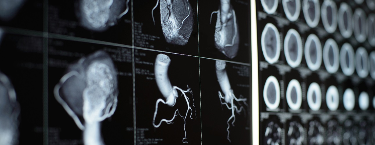
Advanced imaging techniques can display three-dimensional pictures within the arteries, and perhaps provide clues to future cardiovascular events. There are three main types:
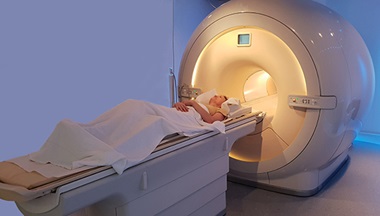
- Coronary calcium scoring/screening – This test uses a CT scan to take an image the heart, which looks for calcium deposits within the coronary arteries. Calcium is not present in normal coronary arteries and when present is a sign of atherosclerosis. A recent NIH study has shown that the more coronary calcium you have, the greater your risk of a heart attack.
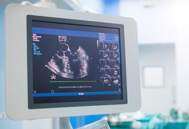
- Carotid ultrasound – A new imaging test that looks for thickness of the inner lining of the carotid arteries. It’s been shown that the more this artery thickens, the higher the risk of cardiovascular disease.
- CT coronary angiography – Shows calcium deposits and possible blockages or narrowings in the blood vessels. A CT coronary angiography is used to determine whether symptoms such as chest pain or shortness of breath are related to a coronary problem, and whether those symptoms can be treated with medicine, with non-invasive techniques, or with surgery.
Who benefits most from new imaging tests?
New imaging tests are a way to assess the cardiovascular condition among patients not necessarily at low or high risk for cardiovascular disease, but in a kind of middle area where the physician needs to determine future diagnosis or treatment.
For healthier patients, that decision can be to keep them on a healthy track. For patients who show signs of borderline-high cardiovascular risk, the physician might recommend modifying their diet or taking cholesterol-lowering medication.
It is among this intermediate-risk group of individuals that new imaging can make a difference. Here are a few of the more important things to remember about the relevance of new imaging:
- When used properly, new imaging can be useful in putting someone on a diet and exercise plan before they need surgical intervention.
- New imaging tests do not replace understanding, recognizing, and managing traditional cardiovascular risk factors such as blood pressure, smoking, diabetes, diet, weight, and exercise.
- New imaging may be useful when deciding the next step in treatment, or whether to start treatment at all. For example, this can be especially relevant for people with heart disease in the family, and who are trying to decide whether their current living habits are healthy enough.
- In non-lifesaving situations, new imaging tests such as CT angiograms can be a simpler alternative to surgical procedures such as stenting or catheterization.
- Women who are post-menopausal have increased cardiovascular risk, and might benefit from new imaging such as calcium screening.
- CT coronary angiography might be useful for older women where there’s a question about the nature of their symptoms. Women often have non-classical symptoms of heart disease, or experience a higher rate of false-positive and false-negative stress tests.
New Imaging and Radiation Risk Among Women
Techniques have recently been introduced that have dramatically lowered radiation exposure. However, CT angiography and other types of computerized tomography still carry some degree of radiation risk. They are not recommended for younger women, especially when used to image the chest. CT scans are also not recommended for pregnant women. As women age, their radiation risk becomes lower, especially in the years after menopause.
Johns Hopkins Women's Cardiovascular Health Center