Hip Dysplasia
What You Need to Know
- Hip dysplasia can run in families. Girls are more likely to have it than boys.
- Babies born in the breech position and firstborn babies are also more likely to be diagnosed with hip dysplasia.
- Hip dysplasia is often diagnosed in babies, but in mild cases with no symptoms, it can go unnoticed until teenage years or adulthood.
- Untreated hip dysplasia can lead to difficulty walking, hip pain and early arthritis.
- Treatments include bracing, casting and/or surgery to promote proper development and stability of the hip joint.
What is hip dysplasia?
Hip dysplasia, sometimes called developmental dysplasia of the hip (DDH) or congenital hip dysplasia, is a range of conditions related to improper hip development or instability of the hip joint. One or both hips may be affected by hip dysplasia.
The hip is a ball-and-socket joint: The ball (the head of the femur) and the socket (acetabulum) rely on each other for development. Hip dysplasia has a spectrum: The mildest form is a shallow socket (acetabulum). The most severe form is a dislocated hip, when the ball is not in the socket. In between, the ball can slide in and out of the socket, which is known as hip subluxation.
Hip dysplasia is commonly diagnosed during infancy or early childhood — usually six to eight weeks after birth, when the hip joint becomes more stable — but it can affect all age groups. It is important that children get a diagnosis and necessary care early in life. If left undiagnosed or unsuccessfully treated, hip dysplasia may lead to early hip arthritis (affecting people as young as their 30s).
On Call for All Kids - Hip Dysplasia in Babies

Risk Factors and Causes of Hip Dysplasia
Hip dysplasia is thought to develop before birth or shortly after. It is often hard to tell when the dysplasia started. There are several genetic and environmental factors that have been linked to the higher risk of developing hip dysplasia. The major risk factors include:
- Being in breech position (feet or bottom first in the uterus) in the third trimester
- Family history of hip dysplasia
- Being swaddled too tightly
- Too little amniotic fluid during pregnancy (oligohydramnios)
Research also shows that hip dysplasia occurs more frequently in babies who are:
- Firstborn children
- Female
- Twins
- High birth weight
Often, there is no single cause of hip dysplasia, and many people diagnosed with hip dysplasia don’t have any of the major risk factors.
Other Causes of Hip Dysplasia
Hip dysplasia can develop alongside genetic disorders such as arthrogryposis, multiple epiphyseal dysplasia, myelomeningocele, Larsen’s syndrome and Ehlers-Danlos syndrome.
It may also accompany neuromuscular disorders such as cerebral palsy and spina bifida. This is called teratologic hip dysplasia, and it often results in a hip dislocation that occurs before birth and usually needs surgery.
Sometimes, hip dysplasia is part of a known birth defect. One example is proximal femoral focal deficiency — a condition in which the ball part of the femur is malformed or missing.
Types of Hip Dysplasia
Hip dysplasia is a spectrum of conditions ranging from malformed hip bones to a dislocated hip:
- Dysplasia: The socket part of the hip joint is shallow or otherwise malformed, but the ball stays within the socket.
- Subluxation: The ball can slide in and out of the hip joint. This can often be noticed on physicial exam with a hip “clunk.” Sometimes, only part of the ball is inside the hip socket.
- Dislocation: The ball part of the joint is fully outside of the socket. A new false socket may form that is not connected to the original socket. If there is no false socket, the ball part is not anchored to a specific part of the hip and can move around within a certain radius of the original socket.
Symptoms of Hip Dysplasia
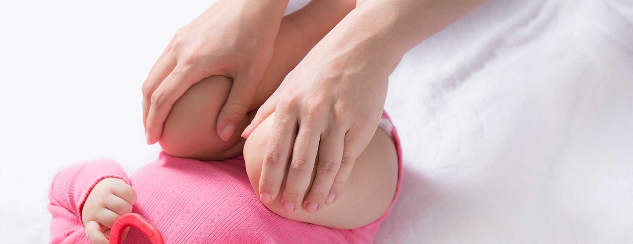
There are certain visual signs that may indicate hip dysplasia in babies when it affects one of the hips:
- Limb length inequality (legs with different lengths)
- Uneven skin folds in the thighs and where buttocks transitions to the thigh
- Abnormal range of motion in one of the hips
A dislocated hip does not typically cause pain in early childhood or prevent a child from learning to walk. Hip dysplasia symptoms in children who can walk include:
- Limited range of motion in the legs
- Abnormal walking or gait, such as limping or toe walking
- Abnormal curvature of the spine (hyperlordosis)
Teens and adults may experience the following symptoms:
- Pain in the hip, back or groin that becomes worse with activity
- Limping
- A catching, snapping or popping sensation in the hip
Hip Dysplasia Diagnosis
If hip dysplasia is severe, it is often caught during a physical exam after birth or during one of the first well-child visits. However, mild hip dysplasia that does not have any visual signs and does not cause symptoms might be diagnosed later in life when the symptoms start.
Diagnostic testing for hip dysplasia may include:
- Physical exam: Patients are evaluated in the clinic using special tests, such as the Barlow test, Ortolani maneuver and Galeazzi sign. These tests help assess the stability and range of motion of the hip joint. When hip dysplasia is present in both hips, it is more difficult to diagnose with a physical exam because the hip movement may appear symmetrical.
- Ultrasound: If hip dysplasia is suspected, infants are referred for an ultrasound in the first six months of life. If a baby has risk factors for hip dysplasia, an ultrasound may be recommended even if the physical exam does not show abnormalities.
- X-ray: An X-ray offers the best visibility into the hip joint, but it cannot provide a clear picture until the hip bones fully solidify, which happens around six months after birth. X-rays are the standard test for older children, teens and adults. They can also be used as secondary imaging for babies who have risk factors and had a normal ultrasound.
Diagnosis in Teens and Adults
Hip dysplasia can be diagnosed for the first time in teens. As the bones grow and mature, mild hip dysplasia that was not caught in infancy may worsen and start causing symptoms. In some cases, hip dysplasia diagnosed and treated in infants may resurface in teens if the treatment was unsuccessful or the progress was not monitored.
When hip dysplasia is diagnosed in teens and adults, it is often called acetabular dysplasia, which refers to the shallow acetabulum (the hip socket).
Hip Dysplasia Treatment
Treatment for hip dysplasia varies depending on the person’s age and the severity of hip dysplasia.
Nonsurgical Treatments
Nonsurgical treatments focus on placing the ball deep into the socket to encourage normal development of the hip. When hip dysplasia is diagnosed early, it can often be treated very successfully with nonsurgical methods.
- Observation: Some infants may outgrow hip dysplasia. Monitoring may be sufficient to see if the condition improves on its own over the first two to six weeks after birth.
- Bracing: Several types of braces are available to help treat hip dysplasia in babies by securing hips in a certain position (like a frog’s legs) that allows for better formation of the hip joint. These braces are typically worn 24/7 for several weeks, although sometimes they may be worn only during sleep or can be taken off for bathing:
- A Pavlik harness is typically recommended for children between six weeks and six months of age. It is a full-body harness that secures around the baby’s shoulders and waist. It is one of the most successful nonsurgical treatments in orthopaedics.
- A rhino brace can be used by children between 3 and 24 months old. It does not extend above the waist, but it restricts movement in the hips. This is sometimes used if a Pavlik harness is not successful.
- Closed reduction and casting: During this procedure, a surgeon places the ball back into the socket and secures it with a lower body cast (spica cast). This is done while the baby is under general anesthesia. Imaging is used to confirm the hip alignment. The cast is worn for several months, and may be changed during that time. Closed reduction and casting may be recommended for children older than 6 months and those who did not improve after bracing. Bracing is used after casting to help reinforce proper hip alignment.
Hip Dysplasia Surgery
Surgery may be necessary for some children and adults who did not improve after nonsurgical treatment or were diagnosed later in life. The surgery may consist of placing the ball into the socket, correcting the position of the ball or socket, or both.
There are several types of surgical techniques to treat hip dysplasia that can be used during a single procedure. The type of technique a surgeon chooses depends on the age and skeletal maturity of the patient, the health of the hip joint, activity goals and other factors.
- Open reduction: If a closed reduction is unsuccessful or the child is older than 12 months, open reduction can be done by making an incision and removing any tissue that is blocking the ball from fitting into the socket.
- Hip tenotomy: If tight tendons are preventing the ball from fitting in the socket securely, the tendons can be lengthened during this procedure to allow for proper hip mechanics.
- Osteotomy: This procedure involves cutting the bones to reshape parts of the hip joint. The specific approach depends on which bones need reshaping. For example:
- The hip socket can be deepened or redirected, depending on the degree of dysplasia and the patient’s age.
- The ball may be rotated or shortened to better fit inside the socket.
If hip dysplasia was not diagnosed and successfully treated early in life, it may lead to hip problems such as hip impingement, hip instability and arthritis. These problems may need to be addressed separately or alongside treating the dysplasia in older teens and adults. Procedures to address them may include the following:
- Hip impingement and labral tears are often treated with arthroscopic surgery.
- Hip instability and acetabular dysplasia in teens and young adults is treated with hip-preserving surgeries such as periacetabular osteotomy.
- Total hip replacement may be necessary for some adults with hip dysplasia who develop osteoarthritis. This is usually the last resort for severe cases.
Recovery and Complications
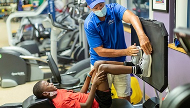
Recovery from surgery may include bracing, casting and physical therapy. Children tend to heal faster than adults. Many patients are limited in their ability to get around for six to eight weeks after surgery. Older patients may need crutches during this period.
Physical therapy, weight-bearing modifications, and follow-up appointments with a doctor are all vital during recovery.
One possible complication after hip dysplasia treatment is proximal femoral growth disturbance (PFGD) of the hip. It occurs when the ball of the hip joint loses blood supply, and the bone starts to die. PFGD is possible after nonsurgical and surgical treatments that force the ball into a position that impairs blood supply.
With treatments such as a Pavlik harness, femoral nerve damage is possible when the hips are spread in such a way that the nerve gets pinched.
Preventing Hip Dysplasia
While it’s not possible to change genetic factors that contribute to hip dysplasia, some environmental factors can be influenced. The main thing parents can do to help prevent hip dysplasia in babies is avoid swaddling. The International Hip Dysplasia Institute offers videos and other resources on safe swaddling techniques if you choose to swaddle.
Medically reviewed by Nakul Talathi, M.D., October 17, 2025