Bladder Cancer Screening and Diagnosis
There are several tests that can determine the diagnosis of bladder cancer. A variety of tests may be used to make an exact diagnosis of bladder cancer.
Pathology Tests
Screening typically begins with pathology tests, where samples of fluid and tissue are examined by a pathologist in a laboratory.
- The most efficient, noninvasive and inexpensive test is a urinalysis/cytology. Here, a sample of urine is taken from the patient and evaluated for cancer cells, red and white blood cells (which fight urinary tract infections), and microscopic hematuria or infection. Hematuria (blood in the urine) is also a sign of a possible urinary tract infection.
- If abnormalities are found in the urine, a biopsy will be performed, in which a pathologist examines tissue for the presence of cancer cells. If the urine culture fails to show abnormalities, a biopsy or other tests still may be ordered — especially if there are symptoms of concern.
Imaging Tests
Imaging tests may be used to locate blockages and tumors, and determine whether cancer has spread to other organs.
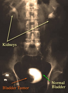
- An intravenous pyelogram is an imaging test during which the patient is injected with dye and the radiologist observes with an X-ray the movement of that dye through the urinary tract. This X-ray will look at the collecting system of the kidneys to determine the presence of any irregularities. This is good for seeing small cancer locations and the upper urinary tract, and especially for detailing the kidneys, ureters and bladder.
- CT or CAT scans are another form of X-ray, which creates a more detailed image of the body and organs. This is used to locate kidney or bladder blockages, and to determine staging, recommended therapy and whether the bladder cancer has metastasized (spread to other parts of the body).
- MRI is another imaging form that creates very high-quality and detailed images of bladder tumors in addition to adjacent organs, such as the chest, pelvis and abdomen, to locate any metastasis.
- Ultrasound imaging, without side effects or radiation, is noninvasive and looks primarily at the bladder and kidneys. It can locate small tract blocks and stones, and also measures the bladder wall thickness.
- The gold standard for the evaluation of the lower urinary tract is a routine outpatient procedure called a cystoscopy. In the same way that a colonoscopy allows doctors to see inside the lower digestive tract, a cystoscopy provides a visual of the lower urinary tract and bladder lining.
During the procedure, a specialized instrument called a cystoscope is passed through the urethra into the bladder. Cystoscopes are either rigid or flexible. Fiber optics allow for images of the bladder lining to be created. If abnormalities, such as tumors, stones or patches of abnormal-appearing tissue, are discovered during cystoscopy, a biopsy may be taken. The biopsy specimen will then be evaluated by a pathologist for the presence of cancerous cells. - Occasionally, the urologist will take a biopsy during a transurethral resection of bladder tumor procedure, which will be scheduled for a future appointment. This is a minimally invasive procedure that does not involve making an incision in the body. The entire removal of a bladder-confined tumor can be accomplished through an operative scope, which passes through the urethra into the bladder.
Bladder Cancer | John's Story
After being diagnosed with bladder cancer by a urologist close to home, John was determined to receive care from an institution with a solid reputation and the most advanced diagnostic and treatment options. This led him to find Trinity Bivalacqua, M.D., Ph.D., of the Johns Hopkins Greenberg Bladder Cancer Institute.