In April, the American Academy of Ophthalmology published a commemorative edition highlighting the seven most influential papers in the journal’s history. Among them was a study by Harry Quigley and W. Richard Green titled “The Histology of Human Glaucoma Cupping and Optic Nerve Damage: Clinicopathologic Correlation in 21 Eyes.” In their introduction to “Ophthalmology: A 2020 Retrospective of Landmark Contributions,” editors Stephen D. McLeod and David W. Parke II called the 1979 article ”a most elegant observational study paired with meticulous histologic evaluation.”
I asked Quigley what it means to say that a study is elegant. It has to have three things, he told me. It has to deal with an idea that no one thought of before, or that changes how everybody thinks. Its design must be flawless, and it has to have incontrovertibly strong evidence for the idea that it’s trying to prove.
To understand the impact of Quigley and Green’s work on the field, it’s helpful to consider the context in which the pair embarked on their study, which looked deeply into the mechanisms of the sight-stealing disease known as glaucoma.
In the late 1970s, experts knew increased eye pressure could lead to glaucoma, and worsen the disease. They knew that the peripheral vision of people with glaucoma deteriorated over time, and that “cupping” — an increase in the size of the optic disc cup relative to the disc — preceded peripheral vision loss. But they didn’t understand the underlying mechanisms of the disease, rendering treatment a guess at best.
This was partly due to a lack of diagnostic tools at the time. Clinicians could get a rough approximation of vision loss through rudimentary manual field tests. They were also able to photograph the inside of the eye, but Quigley says that didn’t tell them what was going on in there. “The most sophisticated piece of equipment we had was the electron microscope, which was good for looking at structure, but not physiology — not how the cells literally behaved,” he says.
Moreover, the only eyes generally available for study were those that pathologists had collected: blind, painful eyes that had been removed — eyes that provided researchers with little insight into what was happening in glaucoma.
Inspired by Ophthalmology Greats
Quigley cites the late Wilmer Eye Institute scientist Jonas Friedenwald, whom he greatly admires, as a big proponent of clinical pathological correlation. That approach involves considering the clinical presentation of a disease — what it looks like in a human — along with microscopic examination of the disease — what’s actually going on to cause the disease. He credits mentor and former Wilmer chairman Edward Maumenee with giving him the confidence to apply clinical pathological correlation to understand what happens in glaucoma.
“One of [Maumenee’s] great passions was talking young men and women who were interested in ophthalmology into thinking they could have a serious influence on the field. He convinced you that you could be the one to do this, and that all you needed was a bit of encouragement,” Quigley says.
Maumenee helped Quigley obtain several stipends to conduct summer research as a resident at Wilmer. One project involved working with Wilmer pathologist Richard Green. “Dick knew every fact about every case of eye disease that had ever been published,” says Quigley. “His approach was to literally take every autopsy eye and eye bank eye that came through the Wilmer Eye Institute and look at every single one of them.”
On Maumenee’s advice, following his residency Quigley did a fellowship at Bascom Palmer Eye Institute. A researcher there was studying axonal transport, the process by which proteins and other organelles are transported between a neuron’s cell body and the nerve endings. Maumenee believed axonal transport had something to do with glaucoma, and this researcher had figured out how to measure axonal transport in the eye. “Maumenee said, ‘You go down there to Bascom Palmer and spend a couple of years learning, and then come back and study that stuff,’” recalls Quigley.
Establishing Cause and Effect
To truly understand what was happening in glaucoma, Quigley knew he needed to study people for whom clinical records and photographs of the eyes were available, and to look at the actual eye itself. During his time at Bascom Palmer, he began collecting eyes. Whenever someone passed away and donated their eyes to the eye bank, Quigley would track down the family and say “I know your family member’s eyes couldn’t be used for corneal transplantation because they had an eye disease, but they can be much more important than that. They can teach us about the eye disease.”
Upon returning to Wilmer, Quigley continued to collect eyes, this time in collaboration with Green. Only now they were collecting the eyes of people who had had glaucoma — and what they saw was very different than what they had been able to see in the blind eyes.
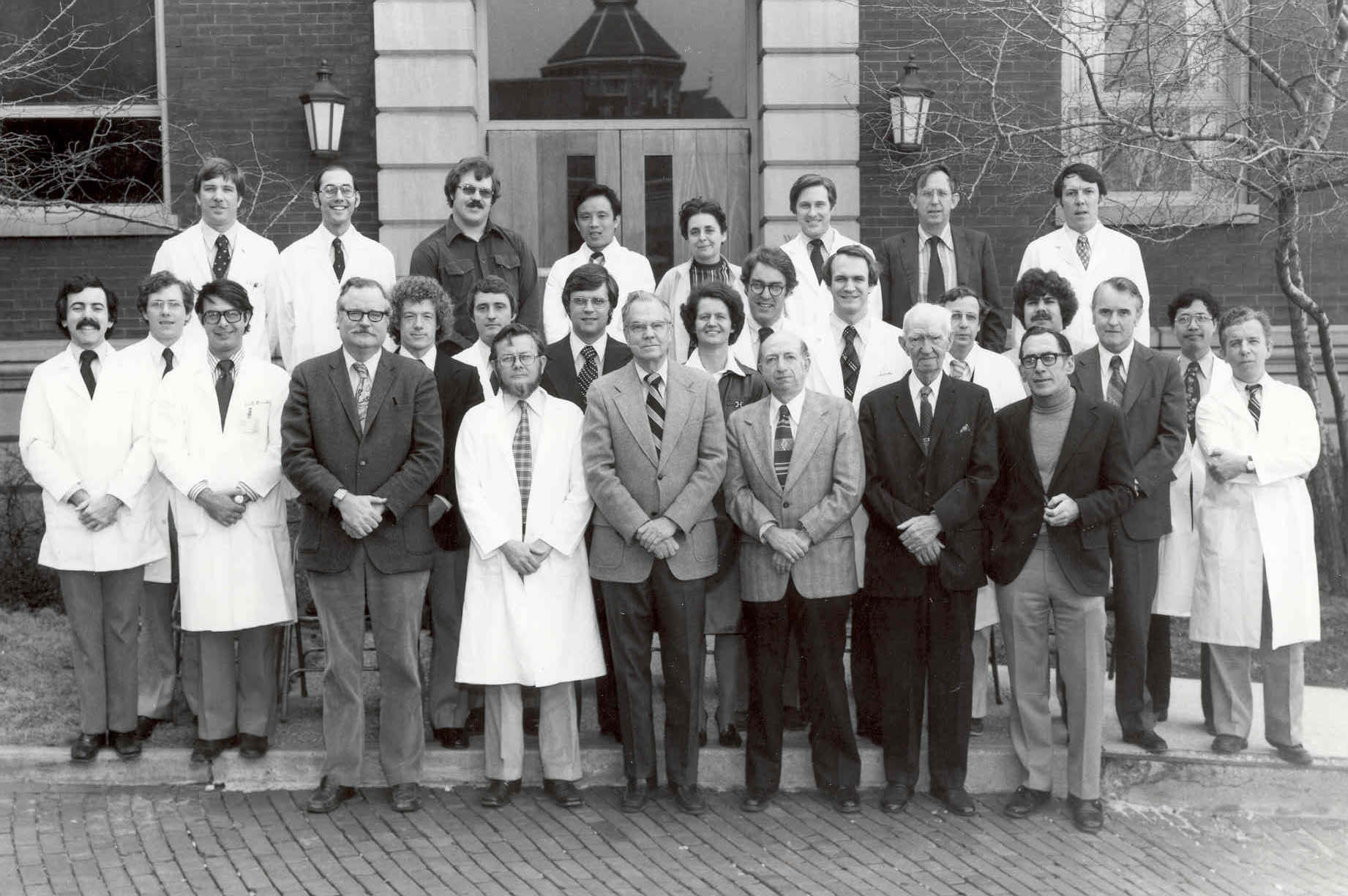 W. Richard Green, front row left in dark jacket, and Harry Quigley, third row second from left, 1978
For their study, Quigley and Green examined 21 eyes of glaucoma patients. Using both standard light microscopy and electron microscopy, they were able to see both the cellular and the connective tissue structures at the back of the eye. For the first time, they determined that when the optic nerve had a cupped appearance, it was because retinal nerve fibers called retinal ganglion cells were dying and causing remodeling of the connective tissues.
They also learned that increased eye pressure was responsible for killing the nerve cells. Importantly, they found that nerve fibers died in significant numbers long before people experienced vision loss, when tests showed that all was well.
Quigley set about finding a way to measure how many nerve cells patients were losing, which would indicate how far advanced the disease was and inform treatment plans. He initially attempted to count the number of nerve cells by using a hand clicker and a felt-tip pen on a printed image of the optic nerve.
Over the next decade, he and Wilmer colleagues Neil Miller and Al Sommer pursued the theory that one could measure the number of dying cells by looking at the nerve fiber layer, an area in the retina where the nerve cell fibers pass down toward the optic nerve. That finding ultimately led to instruments, including optical coherence tomography, used today to measure the number of nerve fibers in the retina.
During this period, the team developed animal models of glaucoma, which further facilitated the study of glaucoma and the development of therapies. Many fellows who worked on those models are now conducting breakthrough research of their own, including the development of gene therapy to block glaucoma from developing.
Building on Sound Science
As with many new ideas, Quigley and Green’s research was initially met with skepticism, but the science behind it was sound and the work was eventually accepted for publication. “I tell my junior colleagues, ‘when you get your work rejected, and when it’s because you’re attempting to change what people think, you’re on the right track,’” says Quigley. He and Green went on to publish a total of nine papers based on their original work.
Quigley says he was greatly privileged in beginning this work and having people provide him with resources and challenge him to go beyond what was known at the time. But he’s quick to add that we still don’t fully understand this disease. “There are lots of diseases, and the translation of what we understand into new therapies is the goal of everything we do.”