Merkin Center Research Grant Recipients
The Merkin Peripheral Neuropathy and Nerve Regeneration Center is proud to announce its latest grants, awarded to nine researchers. Learn more about the recipients and their work below:
Ayobami Ward, M.D., Sc.M.
Resident Physician, Neurosurgery
Post-Doctoral Researcher
University of Michigan
Topic: Age-Dependent Nerve Regeneration Mechanisms: Focus of the Human Repair Schwann Cell Phenotype
-

Ayobami Ward, M.D., Sc.M. is a neurosurgery resident and post-doctoral researcher in the Giger Lab in the Department of Cell and Molecular Biology at the University of Michigan. He received his master’s degree in Biochemistry and Molecular Biology from the Johns Hopkins Bloomberg School of Public Health, before and his Doctorate in Medicine from The Medical College of Georgia. During medical school, his research focused on the neuroimmunology of traumatic brain injury, specifically investigating the role of macrophage polarization in the inflammatory response to cranial trauma.
Currently, his research focuses on the molecular underpinnings of peripheral nerve regeneration with context to human brachial plexus injury and repair. His work utilizes a translational approach to investigating human peripheral nerve injury (PNI) with the goal of improving regeneration and functional outcomes in human patients.
-
The most common forms of nervous system injury are traumatic lesions to peripheral nerves, for which there are few definitive treatments. Although axonal regeneration in the peripheral nervous system (PNS) is more robust than in the central nervous system (CNS), this regeneration is often incomplete and associated with functional deficits. A common form of traumatic PNS injury is damage to the brachial plexus (BP), which is readily observed in newborns after upper extremity traction during birth. In adults, brachial plexus injury is typically observed following trauma to the shoulder or neck. While open surgical BP grafting shows positive functional results in neonates and young children, these same positive benefits are not always seen in adults. The ubiquity of positive outcomes is thought to be limited by multiple factors, however the molecular underpinnings of this delta in functional re-innervation is not well understood.
The proposed study involves a comparative analysis of repair Schwann cell transcriptomes between injured young and aged individuals for the identification of age-dependent, differentially expressed genes (DEGs). Focusing on the injured brachial plexus, we will take human nerve tissue from adult and young patients for molecular analysis and comparison. Our goal is to use these molecular pathways to isolate therapeutic targets and enhance regeneration of PNS/CNS axons after injury. Finally, we hope to translate these discoveries back to patients for enhanced recovery after peripheral nerve injury in the surgical theatre.
Aysel Fisgin, Ph.D.
Research Associate
Johns Hopkins University
Topic: Inhibition of TNIK is a promising therapeutic approach for PIPN
-
Aysel Fisgin, Ph.D., received her BS degree in Chemical Engineering at Middle East Technical University, Turkey, in 1993. After 15 years of working as a professional in industry, then starting up and managing her own business, she started graduate school and got her MS degree in Medical Systems and Informatics from Bogazici University, Turkey. She pursued her Ph.D. in Biomedical Engineering at Bogazici University in Turkey till she moved to the US in 2013. She received a European Union Marie Curie Actions IRSES Project scholarship and carried out her Ph.D. thesis experiments in Biomedical Engineering, Johns Hopkins University. Her Ph.D. thesis research is on high throughput drug screening against CIPN (Chemotherapy-Induced Peripheral Neuropathies). She completed her post-doctoral research fellowship in Hoke Lab, Neurology, Johns Hopkins University and currently works as a research associate in Hoke Lab. She works on in-vitro and invivo peripheral neuropathy models of different chemotherapy drugs and transgenic mice that are resistant to the development of neuropathies caused by these chemotherapy agents. Her research focus is on understanding mechanisms of peripheral neuropathies and identifying therapeutic drugs that can be co- or pre-administered to cancer patients with chemotherapy agents before axonal degeneration starts.
-
Any possible pharmacologic agent that can prevent CIPN will have a great impact on the lives of cancer patients, as it will allow them to have an optimal dose of chemotherapy for prolonged survival and improved quality of life. Furthermore, it can help reduce the significant excess healthcare and resource costs that cancer patients with CIPN face. Studies have linked the serine-threonine kinases MAP4K’s correlate strongly with neurodegeneration. A member of MAP4K family, TRAF2 and NCK interacting kinase (TNIK), is widely expressed across tissues with relatively enriched expression in the brain and it is overexpressed in many types of cancers and it has also been associated with neurological and psychiatric disorders. In our previous studies, we demonstrated several MAP4K inhibitors, that inhibit TNIK as well, provided a robust protection against axonal degeneration in cellular models of Paclitaxel (PTX), Cisplatin (CDDP) and Bortezomib (BTZ) induced peripheral neuropathies. Using a siRNA inhibition approach, we found out that inhibition of TNIK specifically, is enough to prevent PTX induced neurotoxicity in vitro. We hypothesize that; axon degeneration cascades initiated by PTX can be prevented by specific inhibitors of TNIK and I suggest that TNIK inhibitors can be used as an adjunct treatment with Paclitaxel to prevent the onset of peripheral neuropathies. The objectives of this project are; validation of in vitro neuroprotective effect of a specific TNIK inhibitor, with Paclitaxel with regards to axonal degeneration, then to examine the effect of TNIK inhibition at in-vivo model of PTX induced Peripheral Neuropathy with both mice carrying a knock-out mutation in TNIK and a selective inhibitor of TNIK. We will also examine whether inhibition of TNIK will block Paclitaxel’s ability to kill cancer cells.
Jeremy Sullivan, Ph.D.
Assistant Professor
Johns Hopkins University
Topic: Molecular mechanisms of TRPV4-mediated motor axon degeneration
-

Dr. Jeremy Sullivan is an Assistant Professor in the Department of Neurology, Johns Hopkins University School of Medicine. Dr. Sullivan completed his BSc in Biology and his Ph.D. in Neuroscience at the University of Melbourne (Australia). He then undertook postdoctoral fellowships in the laboratories of Dr. Barbara Beltz (Wellesley College, MA), Dr. Pierre Meyrand (CNRS, France), and Dr. Sharon Oleskevich (Garvan Institute of Medical Research, Australia).
Dr. Sullivan joined the laboratory of Professor Charlotte Sumner at Johns Hopkins University in early 2011, where he has been developing and studying both cellular and animal models of TRPV4-mediated hereditary neuromuscular disease. His current research is focused on elucidating the molecular mechanisms via which mutant forms of TRPV4 precipitate neurodegeneration and on developing therapeutic strategies.
-
Blood vessels within nervous tissues exhibit a series of specializations, known as blood-neural barriers (BNBs), which prevent neurons being exposed to neurotoxic blood components. Impairments of BNB function occur in multiple forms of peripheral neuropathy, though little is known about the molecular mechanisms via which leakage from blood triggers disease. In recent work, we have identified the cation channel TRPV4 as a key regulator of BNBs protecting spinal motor neurons. The goals of the proposed studies are to investigate blood components impacting motor axon integrity following BNB breakdown, and to characterize the molecular pathways via which they drive pathology. The findings of these studies will have broad implications for understanding mechanisms underlying motor axon pathology across multiple forms of peripheral neuropathy and for identifying therapeutic strategies promoting axon integrity.
Jesse Stokum, M.D., Ph.D.
Surgery Fellow
Johns Hopkins University
Topic: Enhanced Peripheral Neural Regeneration with Limb Lengthening
-
Jesse Stokum, M.D., Ph.D., is a peripheral nerve surgery fellow working under Dr. Allan Belzberg at the Johns Hopkins Hospital, and a neurosurgery resident physician at the University of Maryland School of Medicine.
He hails from rural southwestern PA and received his M.D. and Ph.D. degrees from The University of Maryland School of Medicine.
-
Peripheral nerve injuries (PNI) occur in 2.8% of civilian trauma patients and 8.1% of combat casualties. The most critical barrier to improved outcomes is the slow rate of nerve regeneration since after injury the denervated distal nerve becomes less hospitable to regenerating nerve fibers and target muscles undergo irreversible atrophy. We recently published our experience regarding a patient with a proximal sciatic nerve transection who showed accelerated nerve regeneration rate after an innovative treatment strategy. To approximate the severed ends of the nerve for direct repair, an 8 cm osteotomy was made to shorten the femur. An expandable intramedullary nail was then used to re-lengthen the femur 1 mm/day. The patient exhibited return of ankle plantarflexion in 9 months, 21 months earlier than expected. If the factor that accelerated our patient’s regeneration can be identified, it could be harnessed to improve recovery after PNI. Here, we propose to establish a clinically relevant large animal model of our patient’s treatment course. The purpose of this model is to (1) replicate and confirm the phenomenon of accelerated nerve regeneration with limb lengthening, and (2) serve as an experimental platform for future studies.
Kathryn Moss, Ph.D.
Research Associate
Johns Hopkins University
Topic: Exploring Functional Demyelination Pathomechanisms for CMT1A and HNPP Caused by Node of Ranvier Defects
-
Dr. Kathryn Moss received her BS degree in Cellular and Molecular Biology from the University of Michigan and her Ph.D. in Biochemistry, Cell and Developmental Biology from Emory University. She completed her dissertation with Dr. Gary Bassell studying posttranscriptional mechanisms of neuronal development and neurological disease. Dr. Moss is currently a postdoctoral fellow in the laboratory of Dr. Ahmet Höke at Johns Hopkins University School of Medicine. Her research interests are focused on understanding the pathogenesis of Charcot-Marie-Tooth disease Type 1A (CMT1A) and Hereditary Neuropathy with Liability to Pressure Palsies (HNPP). Remarkably, these dysmyelinating peripheral neuropathies are caused by copy number variation of the same gene; the Peripheral Myelin Protein 22 (PMP22) gene is heterozygously duplicated in CMT1A and heterozygously deleted in HNPP. Although CMT1A and HNPP are the most common inherited peripheral neuropathies, no disease-modifying treatments are currently available. Dr. Moss is working to understand the pathophysiology of CMT1A and HNPP and identify pathomechanisms causing them to guide therapy development. Dr. Moss’ research endeavors are enhanced by her involvement in the Peripheral Nerve Society. She serves as Chair of the Junior Committee and as a member of several other committees.
-
Charcot-Marie-Tooth Disease Type 1A (CMT1A) and Hereditary Neuropathy with Liability to Pressure Palsies (HNPP) are the most common inherited peripheral neuropathies and even though these diseases dramatically affect patient quality of life and burden the healthcare system, pathomechanisms causing them remain unclear and there are currently no disease-modifying treatments. My previous findings with CMT1A model mice indicate that myelin dysfunction is sufficient to drive functional deficits without inducing secondary axon degeneration and my preliminary results suggest that Node of Ranvier organization is disturbed in peripheral nerve myelin from CMT1A and HNPP model mice. Therefore, I intend to explore functional demyelination as a pathomechanism of CMT1A and HNPP, where myelin sheaths are grossly normal but dysfunctional, by thoroughly evaluating Node of Ranvier organization, development, and maintenance. Results from these studies will provide insight into CMT1A and HNPP pathogenesis and potentially identify targets for developing novel therapeutics.
Sachin Gadani, M.D., Ph.D.
Neuroimmunology Fellow
Johns Hopkins Hospital
Topic: Exploring the role of alarmins on immune recruitment after peripheral nerve injury
-
Sachin Gadani, M.D. Ph.D., is a neurologist-scientist with expertise in neuroimmunology. He earned his Ph.D. at the University of Virginia studying the immune response after injury in the spinal cord. He then went on to complete neurology residency at Johns Hopkins Hospital, and is currently a fellow in the neuroimmunology division.
He has a long-standing interest in understanding how neuroimmune interactions affect recovery from injury. His graduate work focused on ‘alarmins’, signals that initiate inflammation after injury, in the central nervous system. He found that certain alarmins in the brain and spinal cord recruit beneficial cells to the injury site, and improve outcomes.
-
Peripheral nerve injury is a common cause of disabling pain, weakness, and sensory loss. While the peripheral nervous system (PNS) retains the capacity to regenerate, factors such as age, chronicity, and length of denervation prevent adequate regeneration in many people. After PNS injury, Schwann cells are activated into a reparative phenotype, the distal axon becomes fragmented through Wallerian degeneration, and macrophages are recruited to phagocytose myelin and axonal debris alongside Schwann cells. Schwann cells and infiltrating macrophages are critical players in facilitating neuroregeneration; however, the signals that initiate Schwann cell activation and macrophage recruitment in PNS injury remain poorly understood.
In this project, we are exploring the role for alarmins in initiating inflammation after PNS injury, using the murine model of sciatic nerve crush. We speculate that inhibiting alarmins in this model will reduce the recruitment of macrophages into the injured sciatic nerve, and therefore reduce the normal process of nerve regeneration. The long-term goals of this project are to test whether people who have poor or incomplete regeneration have lower or altered levels of alarmins, and whether supplementation with alarmins could improve healing.
Wonjin Yun, Ph.D.
Post-doctoral Research Fellow
Johns Hopkins University
Topic: Modeling CMT1A with a novel macrophage-integrated neural crest organoids (MINOs) and interrogating cell therapy efficacy of hypoimmunogenic induced human Schwann cells.
-
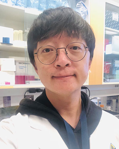
Dr. Wonjin Yun is a postdoctoral research fellow at Johns Hopkins University. His research focuses on unraveling the pathogenic impact of macrophages on CMT1A using CMT1A patient iPSCs-derived organoids. Additionally, he explores the potential of hypoimmunogenic induced Schwann cells for future cell therapy applications.
He received his B.S. and Ph.D. degrees from Korea University, South Korea. During his Ph.D. studies, he delved into understanding the mechanisms of demyelinating diseases and utilizing stem cells for translational research. Specifically, his work centered on generating oligodendrocytes from human pluripotent stem cells through differentiation and from human somatic cells via direct conversion. This research aimed to advance the study of multiple sclerosis by transplanting oligodendrocytes and modeling X-linked adrenoleukodystrophy. In 2021, Dr. Yun joined Johns Hopkins University as a postdoctoral fellow in Dr. Gabsang Lee's laboratory. Since then, he has been dedicated to investigating neurological disorders such as Charcot-Marie-Tooth (CMT), amyotrophic lateral sclerosis (ALS), and scleroderma.
-
Many peripheral neuropathy studies indicate a link between disease manifestation or progression and macrophage recruitment. In fact, significantly elevated numbers of macrophages are found in the affected lesions of CMT1A. Our group previously reported that CMT1A-specific Schwann cells exhibited increased expression of genes involved in chemotaxis, complement activation, and inflammation pathways.
In this study, we will investigate the pathophysiological mechanism of macrophages contributing neuropathy in CMT1A and seek to find the therapeutics for mitigating neuropathy in CMT1A. To achieve this, we will utilize previously established neural crest organoids, differentiated macrophages from CMT1A-hiPSCs, and hypoimmunogenic human induced Schwann cells. To assess pathogenetic impact of macrophages in human CMT1A, we will develop macrophage-integrated neural crest organoids (MINOs) by co-culturing neural crest organoids and differentiated macrophages from CMT1A-hiPSCs followed by detailed molecular characterization, including degree of demyelination. To address peripheral nervous system (PNS) neuropathy in CMT1A, we will transplant hypoimmunogenic human induced Schwann cells into MINOs in vitro, evaluating changes in the degree of CMT1A neuropathy and functional alterations, including de novo myelination.
Xuewei Wang, Ph.D.
Assistant Professor
University of South Florida
Topic: Elucidating the mechanisms by which H3K27me3 maintains normal axon regeneration.
-
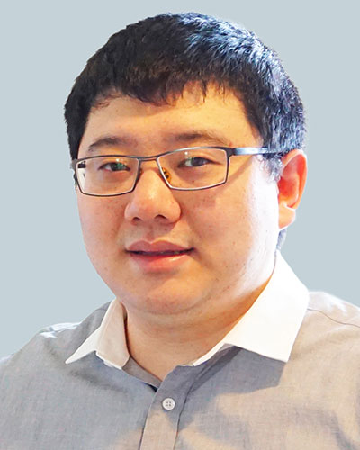
Xuewei Wang, Ph.D. is a neurobiologist who studies molecular mechanisms of mammalian axon regeneration and neuroprotection. Dr. Wang received his Bachelor of Medicine in Basic Medical Sciences and Ph.D. in Neurobiology from Peking University (Beijing, China). He then completed his postdoctoral training at Johns Hopkins University under the supervision of Dr. Fengquan Zhou. He is currently an Assistant Professor at University of South Florida Morsani College of Medicine.
Using mouse sciatic nerve regeneration and optic nerve regeneration models, Dr. Wang’s postdoctoral work focused on two important factors regulating the intrinsic axon regeneration ability of neurons: gene transcription regulation and cytoskeleton dynamics. He recently started his independent career at the current institution. His laboratory will combine multiomics sequencing and axon regeneration models to delineate the epigenomic and transcriptiomic landscapes facilitating axon regeneration.
-
Current clinical treatments for neural injuries and neurodegenerative diseases fall short of success. Accelerating neural repair has been a long-standing challenge for biomedical research. Although neurons in the mammalian peripheral nervous system (PNS) can spontaneously regenerate axons, in humans it is often incomplete, leading to poor functional recovery and permanent disabilities. Therefore, it is critical to develop therapeutics that can accelerate PNS axon regeneration and improve functional recovery in patients.
My published work demonstrates that following peripheral nerve injury, H3K27me3, a histone mark associated with gene transcription suppression, is increased in dorsal root ganglion (DRG) neurons to maintain normal axon regeneration. In this proposed study, I plan to use unbiased multiomics sequencing of DRG neurons purified by fluorescence-activated cell sorting, integrative data analysis, and in vitro and in vivo functional screening of candidate genes to identify key axon regeneration suppressing genes epigenetically silenced by H3K27me3. Successful completion of this study will advance our knowledge of epigenetic regulation of axon regeneration and uncover novel therapeutic targets for accelerating peripheral nerve regeneration.
Yu Su, M.D., Ph.D.
Research Fellow
Johns Hopkins University
Topic: Targeting Glutamate Carboxypeptidase II (GCPII) to Enhance Nerve Remyelination and Recovery Following Peripheral Nerve Injury in Aged Mice
-
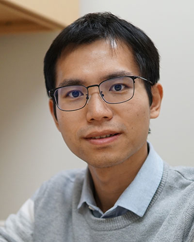
Yu Su is currently a postdoctoral fellow in the Slusher Lab at Johns Hopkins Drug Discovery (JHDD), focusing his research on developing innovative therapeutic interventions targeting the peripheral nervous system (PNS). His investigations center on peripheral nerve regeneration after injury, Schwann cell functionality, and aging-related muscle degeneration.
Dr. Su obtained his M.D., Ph.D. from Central South University in China, undergoing a three-year co-education program with the University of Michigan during his graduate studies. His research experience involved extensive engagement with the PNS, particularly muscle physiology and neuromuscular junctions.
Motivated to bring his PNS experience into clinical translation, Dr. Su joined JHDD—an extraordinary platform facilitating translational research. His current research utilizes small-molecule inhibitors for GCPII enzymes to rejuvenate aged Schwann cells' capacity for remyelination following nerve injury.
-
Peripheral nerve regeneration is a complex process, and despite the innate regenerative capacity of nerves, regeneration is often insufficient, especially during aging. Our project will investigate the role of the enzyme Glutamate Carboxypeptidase II (GCPII) in PNI employing both pharmacological and genetic methodologies. Specifically, we will assess the regulation of GCPII enzyme activity and expression in a murine nerve crush model performed in 22-24 months old mice and the therapeutic benefit of its inhibition using both potent and selective small molecule inhibitors as well as GCPII KO mice.
Our preliminary findings indicate increased GCPII expression and enzymatic activity in the aging peripheral nervous system (PNS), with chronic inhibition demonstrating protection against age-related muscle function decline and neuromuscular junction (NMJ) integrity deterioration. Our preliminary investigation also illustrates enhanced myelination in peripheral nerves with GCPII inhibitor treatment, both in vivo and in vitro. Building on these promising observations, we aim to extend our investigations to assess the role of GCPII in nerve remyelination and recovery following PNIs in aged animals. Success with the proposed studies may lead to novel PNI treatments and facilitate nerve regeneration and remyelination, specifically in the context of aging.
2022 Awardees
- Ashley Kalinski, Ph.D.
Topic: Elucidating the cell-autonomy of SARM1 for injury induced axon regeneration, nerve inflammation, and Schwann cell reprogramming - Atul Rawat, Ph.D.
Topic: Modulating macrophage phenotype in peripheral nerve injury to accelerate nerve regeneration and functional recovery - Baohan Pan, M.D., Ph.D.
Topic: Cutaneous Sensory Innervation in Human and Mouse and Its Implications in Neuropathic Pain - Christopher Cashman, Ph.D.
Topic: Mitochondrial genome mutations and respiratory dysregulation as effectors of diabetic neuropathy - Hyun Sung, Ph.D.
Topic: Deciphering the role of autophagy in the pathogenesis of peripheral neuropathy - Masnsen Cherief, Ph.D.
Topic: Preventing diabetic bone disease using a neuroprotective agent - Pabitra Sahoo, Ph.D.
Topic: Establishing the kinetics for failure of axonal protein synthesis in chronic nerve injury - Qin Zheng, M.D., Ph.D.
Topic: In-vivo characterization of chemotherapy-induced neuropathy using large scale calcium imaging - Simone Thomas, M.S.
Topic: Chemotherapy-Induced Peripheral Neuropathy (CIPN) Assessment
2021 Awardees
- Sarah Berth, M.D., Ph.D.
Topic: Genetic Screen for Axonal Degeneration Modifiers - Aysel Fisgin, Ph.D.
Topic: MAP4K4 Inhibition to Prevent CIPN - Sang-Min Jeon, Ph.D.
Topic: Sprouting Mediated Skin Reinnervation - Ying Liu, M.D., Ph.D.
Topic: Evaluating the effect of SARM1 deficiency on peripheral neuropathy in db/db mouse model of type 2 diabetes. - Brett McCray, M.D., Ph.D.
Topic: TRVP4 in Nerve Injury - Kathryn Moss, Ph.D.
Topic: Development of a CMT1A/CIPN Mouse Model - Bipasha Mukherjee-Clavin, M.D., Ph.D.
Topic: KIF16B-CMT2 - Seong-Hyun Park, Ph.D.
Topic: CMT PNSorganoid Model - Sami Tuffaha, M.D.
Topic: Gene Expression Changes with Schwann Cell Denervation - Eric Villalón Landeros, Ph.D.
Topic: DRG Neuroproteasome Signaling Peptides
