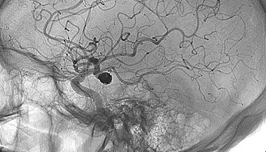
Cerebral Arteriogram
An arteriogram is an X-ray of the blood vessels. It’s used to look for changes in the blood vessels, such as:
- Ballooning of a blood vessel (aneurysm)
- Narrowing of a blood vessel (stenosis)
- Blockages
This test is also called angiogram.
For arteriogram, your healthcare provider inserts a catheter into a large blood vessel and injects contrast dye. The contrast dye causes the blood vessels to appear on the X-ray image. This lets the healthcare provider better see the vessel(s) under exam.
Many arteries can be seen on an arteriogram, including those of the legs, kidneys, brain, and heart. A cerebral arteriogram is used to look at the blood vessels of the brain, head, or neck.
For a cerebral arteriogram, a catheter is usually inserted into an artery in the groin. Sometimes, an artery in the arm is used. Rarely, an artery in the neck may need to be used. The groin artery is most commonly used because it’s easier to get to. Once the catheter is inserted, the contrast dye is injected. Next, a series of X-rays are made. These images show the arteries, veins, and capillaries and blood flow in the brain.
Diagnostic Cerebral Angiography
What is a diagnostic cerebral angiogram?
A diagnostic cerebral angiogram is a medical procedure that helps specialized
doctors examine the blood vessels in the head and neck, including the brain.
These doctors are able to examine blood vessels
using high-tech imaging equipment to take x-ray pictures.
What to expect during your angiogram.
The procedure will take place in a
room like this called the neuroangiography suite.
It was designed specifically to perform procedures like yours.
You'll lie down here with plenty of cushioning
to keep you comfortable while the procedure is done.
Your procedure will be performed under sedation.
This means that your nurse will give you combination of
medications that will help you relax and remove any uncomfortable sensation.
In addition to the sedation medications, the doctor will also apply a
numbing medicine to your groin to ease any discomfort in that area.
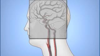
A small tube called a catheter will then be inserted
into an artery in your leg called the femoral artery.
You may feel some pressure as the catheter is being place.
The femoral artery gives the doctor a direct
pathway to the vessels of the head and neck.
Without crossing or coming close to the heart.
How is a cerebral angiogram performed?
The imaging equipment in the neuroangiography sweep uses x-rays to help
the doctors guide the catheter to the vessel they want to see.
When it is time to take the x-ray pictures of the blood vessels,
a contrast medium, or dye is injected into the vessel through the catheter.
This typically produces a warm, but not
painful feeling that lasts for a few seconds.
The imaging equipment may rotate around your head.
You may also hear beeping noises as the pictures are taken.
These noises help to signal your doctor about the position of the equipment.
The X Ray pictures are displayed on screens
that the doctor latch as they're performing the procedure.
The doctor will look at the pictures more closely once your procedure is finished.
What happens after the procedure?
At the end of the procedure, the catheter is removed
and a closure device is used to seal the site.
In selected situations the doctor holds pressure
at that site for 15 to 20 minutes.
A band-aid is then applied.
You will stay here in the recovery room for five to six hours after the procedure.
Thank you very much for watching.
Again, please don't hesitate to share any questions or concerns you have with us.
We hope your experience at Johns Hopkins
is as pleasant and comfortable as possible.
Why might I need a cerebral arteriogram?
This test may be advised when previous tests don’t give enough information.
A cerebral arteriogram is used to look for changes in the blood vessels within or leading to the brain. Such as:
- Ballooning or bulging of a blood vessel (aneurysm)
- Blood vessel narrowing (stenosis)
- Narrowing of the arteries (atherosclerosis)
- Inflammation of the blood vessels that narrows them (vasculitis)
- Abnormal connection between the arteries and veins (arteriovenous malformation)
- A blood clot within a blood vessel (thrombosis)
- A spasm of the blood vessel causing an irregular narrowing of the vessel (vasospasm)
- Complete blockage of a blood vessel
Conditions that cause a displacement of the brain's blood vessels may also be seen. These conditions include:
- Tumors
- Stroke
- Swelling (edema)
- Dislocation of the brain tissue, caused by pressure within the brain due to swelling, bleeding, or other reasons (herniation)
- A tear in an artery
- Increased pressure within the brain
- Fluid in the brain (hydrocephalus)
A cerebral arteriogram may be used to locate or assess clips on blood vessels placed during previous surgical procedures.
There may be other reasons for your healthcare provider to recommend a cerebral arteriogram.
What are the risks of cerebral arteriograms?
You may want to ask your healthcare provider about the how much radiation is used during the procedure and the risks related to your situation. It’s a good idea to keep a record of your history of radiation exposure, such as previous CT scans and other types of X-rays, so that you can tell your healthcare provider. Risks associated with radiation exposure may be related to the number of X-rays and/or treatments over a long period.
If you are pregnant or think you may be, tell your healthcare provider. Radiation exposure during pregnancy may lead to birth defects. If you need to have a cerebral arteriogram, special precautions will be taken to reduce the radiation exposure to the fetus.
There is a risk for an allergic reaction to the dye used for this test. If you are allergic to or sensitive to medicines, contrast dye, or iodine, tell your healthcare provider. Also, tell your healthcare provider if you have kidney failure or other kidney problems.
Tell your healthcare provider if you have liver or thyroid conditions. In some cases, this procedure is not advised for people with these conditions.
Tell your healthcare provider if you have a history of bleeding disorders or if you are taking any anticoagulant (blood-thinning) medicines, aspirin, or other medicines that affect blood clotting. You may need to stop these medicines before the procedure.
Because the procedure involves the blood vessels and blood flow of the brain, there is a small risk for complications involving the brain. These complications may include:
- Loss of consciousness
- Transient ischemic attack (TIA, a brief stroke-like condition)
- Paralysis on one side of the body (hemiplegia)
- Blood clot in the blood vessel (embolus)
- Bleeding
- A collection of blood and swelling (hematoma)
- Stroke
- Loss of the ability to speak or understand speech (aphasia)
There may be other risks depending on your specific medical condition. Be sure to discuss any concerns with your healthcare provider before the procedure.
How do I prepare for cerebral arteriogram?
- Your healthcare provider will explain the procedure to you and ask if you have questions.
- You will be asked to sign a consent form that gives permission to do the procedure. Read the form carefully and ask questions if anything is not clear.
- Be sure to tell your healthcare provider, the radiologist, or the technologist if you have ever had a reaction to any contrast dye, or if you are allergic to iodine or shellfish.
- Tell your healthcare provider if you are sensitive to or are allergic to any medicines, latex, tape, and anesthetic agents (local and general).
- You will need to fast (not eat) for a certain time prior to the procedure. Your healthcare provider will tell you how long to fast, whether for a few hours or overnight.
- Tell your healthcare provider if you are pregnant or think you may be.
- Make sure your healthcare provider has a list of all medicines (prescribed and over-the-counter) and all herbs, vitamins, and supplements that you are taking.
- You may be given a medicine to relax you and make you sleepy before the procedure. You may also receive an anticholinergic medicine, which slows down the production of saliva in the mouth, blocks the production of acid in the stomach, and slows down the activities of the intestinal tract, among other effects. If you get this medicine, you may notice that your mouth feels dry.
- Tell your healthcare provider if you have a history of bleeding disorders or if you are taking any anticoagulant (blood-thinning) medicines, aspirin, or other medicines that affect blood clotting. You may need to stop these medicines before the procedure.
- Depending on the site used for injection of the contrast dye, the recovery period may last up to 12 to 24 hours. You should be ready to spend the night if necessary.
- Your healthcare provider may request a blood test prior to the procedure to see how long it takes your blood to clot. Other blood tests may be done as well.
- Based on your medical condition, your healthcare provider may give you other instructions on what to do before the procedure.
How is the cerebral arteriogram done?
A cerebral arteriogram may be done on an outpatient basis or as part of your stay in a hospital. Procedures may vary depending on your condition and your healthcare provider's practices.
Generally, a cerebral arteriogram follows this process:
- You will be asked to remove any clothing, jewelry, hairpins, dentures, or other objects that may get in the way of the procedure.
- If you are asked to remove clothing, you will be given a gown to wear.
- You will be reminded to empty your bladder before starting the procedure, which can take up to 3 hours.
- You will be positioned on the X-ray table.
- You may be connected to an electrocardiogram (ECG) monitor that records the electrical activity of your heart. Your vital signs (heart rate, blood pressure, and breathing rate) and neurological signs will be watched during the procedure.
- The catheter (a thin, soft tube) will be put into an artery in either your neck, arm, or groin after the skin is cleansed and a local anesthetic (numbing medicine) is injected.
- If the catheter will be put into an artery in your groin or the arm, the radiologist will check your pulses below the site and mark them with a marker so that the circulation to the limb below the site can be checked after the procedure. In some cases, the catheter is put into an artery in your neck. If the neck is used, a pillow will be placed under your shoulders to keep your neck extended. Your head will be held in place with a strap or tape to prevent the risk of damage to the artery that might happen if you move your head. If the groin or arm is used, the site will be shaved prior to insertion of the catheter. If the arm is used, a blood pressure cuff will be applied to your arm below the insertion site and inflated to prevent flow of the contrast dye into your lower arm.
- Once the catheter is put into the artery at the groin or arm, it’s threaded through to an artery in the neck. A special type of X-ray, called fluoroscopy, may be used to verify the location of the catheter inside your body.
- An injection of contrast dye will be given. The contrast dye makes the blood vessels show up on the X-ray image. This allows the healthcare provider to better see the structure of the vessel(s).You may feel some effects when the dye is injected into the catheter. These effects include a flushing sensation, a salty or metallic taste in your mouth, a brief headache, or nausea and/or vomiting. These effects usually only last for a few moments.
- You should tell the radiologist right away if you have any breathing difficulties, sweating, numbness, or heart palpitations.
- After the contrast dye is injected, a series of X-rays will be taken. The first series of X-rays shows the arteries, and the second series shows capillary and vein blood flow.
- Depending on the study being done, there may be one or more injections of contrast dye.
- When the test is done, the catheter will be removed and pressure will be applied over the area to keep the artery from bleeding.
- After the site stops bleeding, a dressing will be applied to the site. A sandbag or other heavy item may be placed over the site to prevent further bleeding or the formation of a hematoma at the site.
What happens after cerebral arteriogram?
Depending on which site was used for injection of the contrast dye, you will stay flat in bed in a recovery room for several hours after the procedure. If the groin or arm site was used, the leg or arm on that side will be kept straight for up to 12 hours. If the neck was used, you will be watched for signs of hoarseness, breathing problems, or pain or difficulty in swallowing.
A nurse will monitor your vital signs, your neurological signs, and the injection site while you are in the recovery room.
You may be given pain medicine for pain or discomfort related to the injection site or pain from having to lie flat and still for a long period.
You will be encouraged to drink water and other fluids to help flush the contrast dye from your body.
You may go back to your usual diet and activities after the procedure, unless your healthcare provider tells you otherwise.
When you have completed the recovery period, you may be returned to your hospital room or discharged to your home. If this procedure was done as an outpatient, plan to have another person drive you home.
Home care
Once home, check the injection site for bleeding. A small bruise is normal, as is an occasional drop of blood at the site.
If the groin or arm was used, you should monitor the leg or arm for changes in temperature or color, pain, numbness, tingling, or loss of function of the limb.
Drink plenty of fluids to prevent dehydration and to help flush out the contrast dye.
You may be advised not to do any strenuous activities or take a hot bath or shower for a period after the procedure.
When should I call my healthcare provider?
Get prompt medical attention if any of the following happen:
- Fever and/or chills
- Increased pain, redness, swelling, bleeding, or other drainage from the injection site
- Coolness, numbness and/or tingling or other changes in the affected extremity
- Speech or vision changes
- Dizziness
- Muscle weakness or numbness
- Chest pain
Your healthcare provider may give you other instructions after the procedure, depending on your situation.