Exams We Offer: Bone Densitometry (DEXA)
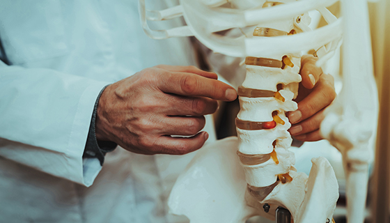
A bone density scan, also called DEXA, uses an X-ray to measure the bone mineral content and density. A DEXA scan produces more detailed images than a standard X-ray and helps to identify fragile bones before they break. A DEXA measures the bone density of spine, pelvis, lower arm and thigh.
Our team at Johns Hopkins Medical Imaging provides compassionate patient care and high-quality imaging in convenient suburban locations. Using the most advanced technology and equipment, our highly trained radiologists and imaging technologists tailor each exam to your specific needs and diagnosis.
Request An Appointment Request An Appointment
Schedule by phone

How do I prepare for a DEXA exam?
-
PRECAUTIONS: Notify the technologist if you are pregnant or think you may be pregnant. Ensure that you have not had a barium or nuclear medicine exam within the past 72 hours.
EAT/DRINK: Do not take calcium supplements 24 hours prior to your exam.
CLOTHING: In some cases, you may stay dressed but will be asked to remove all metallic objects such as belt buckles, zippers, coins, keys and jewelry. In other cases, you will be given a gown to wear so that no buttons, zippers or hooks will interfere with the imaging process.
Based on your medical condition, your health care provider may request other specific preparations.
-
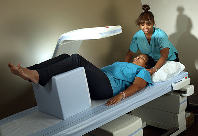
- You will be positioned on a table, lying flat on your back. Your legs will be supported on a padded box which serves to flatten the pelvis and lumbar spine.
- Under the table, a photon generator will pass slowly beneath you, while an X-ray detector camera will pass above the table parallel to the photon generator beneath, projecting images of the lumbar spine and hip bones onto a computer monitor.
- After the scan of the lumbar spine and hip bones is complete, your foot will be inserted into a brace that moves your non-dominant hip (the side you use the least) into a rotated position.
- The next imaging procedure may involve the radius, one of the two bones of the lower arm. The non-dominant arm (the arm you use the least) is usually examined, unless there is a history of a fracture of that arm.
- The computer will calculate the amount of photons that are not absorbed by the bones to determine the bone mineral content. The bone mineral density will then be calculated by a radiologist.
-
- There is typically no special type of care after a DEXA scan. You may resume your normal diet and activities, unless your doctor advises you differently. Your doctor may give you additional or alternate instructions after the procedure, depending on your particular situation.
Who should consider a DEXA scan?
- Women aged 65 and older
- Women discontinuing estrogen should be considered for bone density testing according to all indications listed below.
- For women during the menopausal transition with clinical risk factors for fracture, such as
- low body weight
- prior fracture
- high-risk medication use
- For post-menopausal women younger than age 65, a bone density test is indicated if they have a risk factor for low bone mass such as:
- Low body weight
- Prior fracture
- High risk medication use
- Disease or condition associated with bone loss
- Men aged 70 and older
- For men younger than age 70, a bone density test is indicated if they have a risk factor for low bone mass such as:
- Low body weight
- Prior fracture
- High risk medication use
- Disease or condition associated with bone loss
- Adults with a fragility fracture
- Adults with a disease or condition associated with low bone mass or bone loss
- Adults taking medications associated with low bone mass or bone loss
- Anyone being considered for pharmacologic therapy
- Anyone being treated, to monitor treatment effect
- Anyone not receiving therapy in whom evidence of bone loss would lead to treatment
Why Choose Johns Hopkins Medical Imaging?
Physician Experts

Top Radiology Department
State-of-the-Art Technology

Your Safety Is Always Our Priority

Specialty Technologists

Patient Care
