Kidney Ultrasound
What is a kidney ultrasound?
A kidney ultrasound is a noninvasive diagnostic exam that produces images, which are used to assess the size, shape, and location of the kidneys. Ultrasound may also be used to assess blood flow to the kidneys.
Ultrasound uses a transducer that sends out ultrasound waves at a frequency too high to be heard. The ultrasound transducer is placed on the skin, and the ultrasound waves move through the body to the organs and structures within. The sound waves bounce off the organs like an echo and return to the transducer. The transducer processes the reflected waves, which are then converted by a computer into an image of the organs or tissues being examined.
The sound waves travel at different speeds depending on the type of tissue encountered - fastest through bone tissue and slowest through air. The speed at which the sound waves are returned to the transducer, as well as how much of the sound wave returns, is translated by the transducer as different types of tissue.
An ultrasound gel is placed on the transducer and the skin to allow for smooth movement of the transducer over the skin and to eliminate air between the skin and the transducer for the best sound conduction.
Another type of ultrasound is Doppler ultrasound, sometimes called a duplex study, used to show the speed and direction of blood flow within the chest. Unlike a standard ultrasound, some sound waves during the Doppler exam are audible.
Ultrasound may be safely used during pregnancy or in the presence of allergies to contrast dye, because no radiation or contrast dyes are used.
Other related procedures that may be performed to evaluate the kidneys include X-ray , computed tomography (CT scan) , kidney scan , antegrade pyelogram , intravenous pyelogram , and kidney angiogram .
.png?h=198&iar=0&mh=360&mw=520&w=200&hash=9615D86AF4BE840950C8DC5B0B73C4AC)
How do the kidneys work?
The body takes nutrients from food and converts them to energy. After the body has taken the food that it needs, waste products are left behind in the bowel and in the blood.
The kidneys and urinary system keep chemicals, such as potassium and sodium, and water in balance, and remove a type of waste, called urea, from the blood. Urea is produced when foods containing protein, such as meat, poultry, and certain vegetables, are broken down in the body. Urea is carried in the bloodstream to the kidneys.
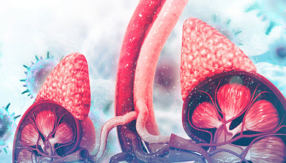
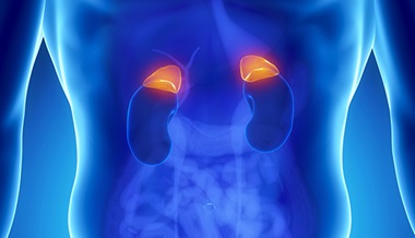
Two kidneys, a pair of purplish-brown organs, are located below the ribs toward the middle of the back. Their function is to:
-
Remove liquid waste from the blood in the form of urine
-
Keep a stable balance of salts and other substances in the blood
-
Produce erythropoietin, a hormone that aids the formation of red blood cells
-
Regulate blood pressure
The kidneys remove urea from the blood through tiny filtering units called nephrons. Each nephron consists of a ball formed of small blood capillaries, called a glomerulus, and a small tube called a renal tubule.
Urea, together with water and other waste substances, forms the urine as it passes through the nephrons and down the renal tubules of the kidney.
What are the reasons for a kidney ultrasound?
A kidney ultrasound may be used to assess the size, location, and shape of the kidneys and related structures, such as the ureters and bladder. Ultrasound can detect cysts, tumors, abscesses, obstructions, fluid collection, and infection within or around the kidneys. Calculi (stones) of the kidneys and ureters may be detected by ultrasound.
A kidney ultrasound may be performed to assist in placement of needles used to biopsy (obtain a tissue sample) the kidneys , to drain fluid from a cyst or abscess, or to place a drainage tube. This procedure may also be used to determine blood flow to the kidneys through the renal arteries and veins.
Kidney ultrasound may be used after a kidney transplant to evaluate the transplanted kidney.
There may be other reasons for your physician to recommend a kidney ultrasound.
Our Approach to Kidney Ultrasounds

The Johns Hopkins Kidney Program is one of the first in the country and our doctors have pioneered some of the most innovative treatments for patients with renal failure. In addition to offering leading-edge procedures, our program offers shorter wait times and minimally invasive options, which can lead to a faster recovery.
What are the risks of a kidney ultrasound?
There is no radiation used and generally no discomfort from the application of the ultrasound transducer to the skin.
There may be risks depending upon your specific medical condition. Be sure to discuss any concerns with your physician prior to the procedure.
Certain factors or conditions may interfere with the results of the test. These include, but are not limited to, the following:
-
Severe obesity
-
Barium within the intestines from a recent barium procedure
How do I prepare for a kidney ultrasound?
EAT/DRINK: Drink a minimum of 24 ounces of clear fluid at least one hour before your appointment. Do not empty your bladder prior to the procedure. Generally, no prior preparation, such as fasting or sedation, is required.
Your physician will explain the procedure to you and offer you the opportunity to ask any questions that you might have about the procedure.
You may be asked to sign a consent form that gives your permission to do the procedure. Read the form carefully and ask questions if something is not clear.
Based upon your medical condition, your physician may request other specific preparation.
What happens during a kidney ultrasound?
A kidney ultrasound may be performed on an outpatient basis or as part of your stay in a hospital. Although each facility may have different protocols in place, generally an ultrasound procedure follows this process:
-
You will be asked to remove any clothing, jewelry, or other objects that may interfere with the scan.
-
If asked to remove clothing, you will be given a gown to wear.
-
You will lie on an examination table on your stomach.
-
Ultrasound gel is placed on the area of the body that will undergo the ultrasound examination.
-
Using a transducer, a device that sends out the ultrasound waves, the ultrasound wave will be sent through that patient's body.
-
The sound will be reflected off structures inside the body, and the ultrasound machine will analyze the information from the sound waves.
-
The ultrasound machine will create images of these structures on a monitor. These images will be stored digitally.
-
If the bladder is examined, you will be asked to empty your bladder after scans of the full bladder have been completed. Additional scans will be made of the empty bladder.
There are no confirmed adverse biological effects on patients or instrument operators caused by exposures to ultrasound at the intensity levels used in diagnostic ultrasound.
While the kidney ultrasound procedure itself causes no pain, having to lie still for the length of the procedure may cause slight discomfort, and the clear gel will feel cool and wet. The technologist will use all possible comfort measures and complete the procedure as quickly as possible to minimize any discomfort.
What happens after a kidney ultrasound?
There is no special type of care required after a kidney ultrasound. You may resume your usual diet and activities unless your physician advises you differently.
Your physician may give you additional or alternate instructions after the procedure, depending on your particular situation.