Anatomy of the Urinary System
How does the urinary system work?
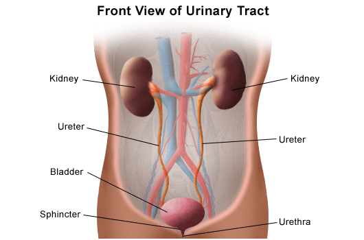
The urinary system's function is to filter blood and create urine as a waste by-product. The organs of the urinary system include the kidneys, renal pelvis, ureters, bladder and urethra.
The body takes nutrients from food and converts them to energy. After the body has taken the food components that it needs, waste products are left behind in the bowel and in the blood.
The kidney and urinary systems help the body to eliminate liquid waste called urea, and to keep chemicals, such as potassium and sodium, and water in balance. Urea is produced when foods containing protein, such as meat, poultry, and certain vegetables, are broken down in the body. Urea is carried in the bloodstream to the kidneys, where it is removed along with water and other wastes in the form of urine.
Other important functions of the kidneys include blood pressure regulation and the production of erythropoietin, which controls red blood cell production in the bone marrow. Kidneys also regulate the acid-base balance and conserve fluids.
Kidney and urinary system parts and their functions
-
Two kidneys. This pair of purplish-brown organs is located below the ribs toward the middle of the back. Their function is to:
-
Remove waste products and drugs from the body
-
Balance the body's fluids
-
Release hormones to regulate blood pressure
-
Control production of red blood cells
-
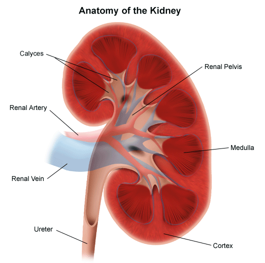
The kidneys remove urea from the blood through tiny filtering units called nephrons. Each nephron consists of a ball formed of small blood capillaries, called a glomerulus, and a small tube called a renal tubule. Urea, together with water and other waste substances, forms the urine as it passes through the nephrons and down the renal tubules of the kidney.
-
Two ureters. These narrow tubes carry urine from the kidneys to the bladder. Muscles in the ureter walls continually tighten and relax forcing urine downward, away from the kidneys. If urine backs up, or is allowed to stand still, a kidney infection can develop. About every 10 to 15 seconds, small amounts of urine are emptied into the bladder from the ureters.
-
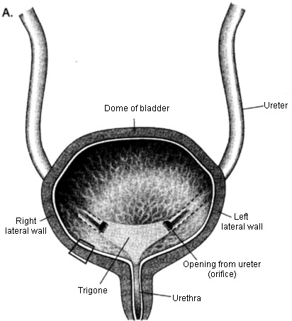
Bladder. This triangle-shaped, hollow organ is located in the lower abdomen. It is held in place by ligaments that are attached to other organs and the pelvic bones. The bladder's walls relax and expand to store urine, and contract and flatten to empty urine through the urethra. The typical healthy adult bladder can store up to two cups of urine for two to five hours.
Upon examination, specific "landmarks" are used to describe the location of any irregularities in the bladder. These are:
-
Trigone: a triangle-shaped region near the junction of the urethra and the bladder
-
Right and left lateral walls: walls on either side of the trigone
-
Posterior wall: back wall
-
Dome: roof of the bladder
-
-
Two sphincter muscles. These circular muscles help keep urine from leaking by closing tightly like a rubber band around the opening of the bladder.
-
Nerves in the bladder. The nerves alert a person when it is time to urinate, or empty the bladder.
-
Urethra. This tube allows urine to pass outside the body. The brain signals the bladder muscles to tighten, which squeezes urine out of the bladder. At the same time, the brain signals the sphincter muscles to relax to let urine exit the bladder through the urethra. When all the signals occur in the correct order, normal urination occurs.
Facts about urine
-
Normal, healthy urine is a pale straw or transparent yellow color.
-
Darker yellow or honey colored urine means you need more water.
-
A darker, brownish color may indicate a liver problem or severe dehydration.
-
Pinkish or red urine may mean blood in the urine.