Sialendoscopy
Featured Expert
Sialendoscopy is a minimally invasive procedure to diagnose and treat salivary gland disorders including stones, strictures, chronic inflammation and other problems affecting the major salivary glands. Doctors use a tiny endoscope to see and address problems inside the major salivary glands.
What You Need to Know
- Sialendoscopy was first practiced in the U.S. in 2004, and head and neck surgeon David Eisele at Johns Hopkins was among the first in the country to use the technique.
- The procedure is an alternative to open surgery on salivary glands, which can cause scarring and complications such as facial nerve damage.
What are salivary glands?
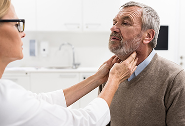
Two pairs of glands on the sides of your face supply saliva to your mouth and keep it moistened and healthy so you can speak, chew and swallow.
The parotid salivary glands are just above the angle of your jaw and in front of your ears. The submandibular salivary glands are under your jaw.
Each of the four glands has an opening, or duct, where saliva flows out. If the duct becomes blocked by a salivary stone or the opening becomes too tight (a stricture), the saliva can back up in the gland, causing swelling, inflammation, pain and infection.
Sialendoscopy can address all these problems, and with less risk of complications than traditional open salivary gland surgery.
What is sialendoscopy?
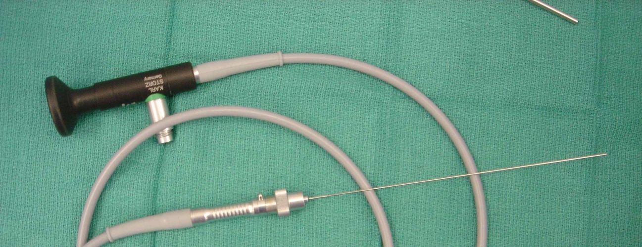
Sialendoscopy is an endoscopic technique to diagnose and treat salivary gland problems. A specialized, very narrow endoscope called a micro-endoscope provides a lighted view of the inside of the gland. The micro-endoscopes range from 0.8 to 1.6 millimeters in width.This micro-endoscope is outfitted with a light, a camera and tools. Tiny baskets and small graspers can extract very small salivary stones. Other tools can help the doctor dilate blockages such as strictures of the salivary duct.“Patients should look for surgeons who have experience in surgical armamentarium and in using the tools that enable more minimally-invasive and gland preserving procedures,” Eisele says.
Sialendoscopy — Why It’s Performed
A doctor may recommend sialendoscopy to address one or more of the following problems:
Salivary Stones
A salivary stone (also called a sialolith) is a hard nugget of mineral deposit that forms in a salivary gland. It can block the duct and cause pain, swelling and infection. Sialendoscopy can aid in the removal of a salivary stone.
Radiation-Induced Salivary Gland Damage
When a person is treated with radioactive iodine to address thyroid gland disease, the salivary glands can take up the radioactive iodine and be damaged by it. In some people, this inflammation ― radiosialadenditis ― is hard to resolve.
Eisele explains, “Patients can experience dry mouth and trouble eating, because the glands don't produce enough saliva. Narrowed ducts can cause stagnation of saliva. Sialendoscopy can dilate ducts and flush out debris, which gives most patients symptomatic relief.”
Chronic Salivary Gland Infection Sialendoscopy can also help manage a chronically inflamed salivary gland (sialadenitis) by dilating the ducts and irrigating the gland.
Sialendoscopy — What to Expect
- Before performing a sialendoscopy, the doctor may order imaging studies such as a computerized tomography (CT) scan, magnetic resonance imaging (MRI) or ultrasound to look at the salivary gland that is causing problems.
- You will receive general anesthesia. Follow your doctor’s instructions on eating, drinking and taking your regular medications before the sialendoscopy.
- The doctor will guide the micro-endoscope into the duct of the salivary gland in your mouth. If the opening is too tight, it may be gently stretched to accommodate the scope.
- The doctor will use the micro-endoscope to rinse (irrigate) the inside of the gland with saline to remove debris.
- Using the light, camera and tools with the micro-endoscope, the surgeon inspects the interior of the gland, and then removes salivary stones and addresses blockages.
- In some cases, the doctor may recommend a sialendoscopy together with traditional open surgery to provide a better view and gain access to the inside of the salivary gland.
Sialendoscopy Risks and Complications
Because sialendoscopy is minimally invasive, there may be less risk of complications than with traditional, open surgery on the salivary gland. However, though rare, sialendoscopy complications are possible and include:
- Strictures (tightening of the duct)
- Perforation of the duct
- Bleeding
- Ranulas, which are benign cysts filled with saliva
After Your Sialendoscopy
After the procedure, you should rest and recover for the remainder of the day. Your salivary glands may be swollen and sore. You may be instructed to take an over-the-counter anti-inflammatory medicine, or you may get a prescription for an alternative.
Complete recovery after a sialendoscopy procedure usually takes only a week or so. Follow your doctor’s instructions, which may include avoiding any heavy lifting or straining.
Ask your doctor about which foods may be easier to eat. You may stick to a soft food diet for a few days after your procedure. Be sure to carefully rinse out your mouth with water after eating to keep food particles out of the treatment area.
Salivary Gland Center
The Johns Hopkins Salivary Gland Center connects you with state-of-the-art evaluation, diagnosis and surgical treatment, including minimally invasive approaches such as sialendoscopy.