CT Scan Versus MRI Versus X-Ray: What Type of Imaging Do I Need?
Featured Expert

Updated July 16, 2026
If you’ve ever had an injury, chances are you’ve had an imaging exam. Imaging tests are extremely powerful tools that can help doctors diagnose a range of conditions. However, imaging tests are not the same as one another. Laura Fayad, M.D., M.S., chief of musculoskeletal imaging at Johns Hopkins Medicine, explains the differences between a CT scan, MRI and X-ray so you can have an informed discussion with your doctor about which type of imaging is right for you.
Key Points
- CT scans, MRIs and X‑rays all visualize internal body structures, but differ in accessibility, detail and the type of energy used.
- X‑rays are fast and widely available, making them ideal for detecting fractures, dislocations and major bone issues, but some types of abnormalities are not visible by X-ray.
- CT scans offer rapid, highly detailed 3D imaging, useful in trauma cases to identify fractures, blood clots and organ injuries, and particularly good at detailing complex anatomy.
- MRIs provide the highest soft‑tissue detail, helping diagnose ligament, tendon, nerve and cartilage injuries without using radiation. At Johns Hopkins, we also use highly detailed imaging (3D imaging) that enables us to see very small structures in joints, bones and soft tissues.
MRIs, CT scans, and X-rays are all diagnostic tools that allow doctors to see the internal structures of the body. They create images using various forms of electromagnetic energy such as radio waves and X-rays. These imaging technologies differ widely when it comes to:
- Accessibility
- Resolution (level of detail in the images)
- Type of energy used
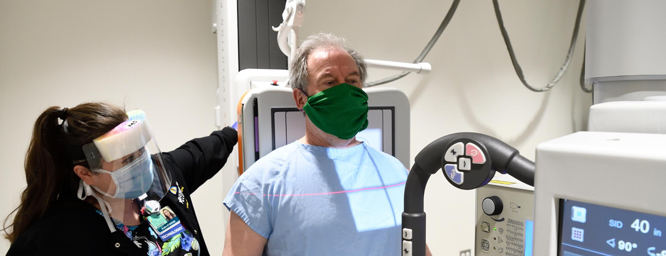
What injuries require an X-ray?
An X-ray, also called a radiograph, sends radiation through the body. Areas with high levels of calcium (bones and teeth) block the radiation, causing them to appear white on the image. Soft tissues allow the radiation to pass through. They appear gray or black on the image.
An X-ray is the fastest and most accessible form of imaging. An X-ray exam only takes a few minutes to complete. “That’s usually the first-line imaging,” explains Fayad. “X-rays often allow us to see major problems with the bones.”
X-rays are ideal for spotting:
- Fractures
- Dislocations
- Misalignments
- Narrowed joint spaces
- Problems with prosthetic hardware
However, an X-ray won’t show subtle bone injuries, soft tissue injuries or inflammation. Even if your doctor suspects a soft tissue injury like a tendon tear, an X-ray might be ordered to rule out a fracture and make sure a bone abnormality is not the reason for symptoms.
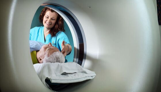
What injuries require an MRI?
An MRI, or magnetic resonance imaging, uses a powerful magnet to pass radio waves through the body. Protons in the body react to the energy and create highly detailed pictures of the body’s structures, including the soft tissues, nerves and blood vessels. Unlike X-rays and CT scans, MRIs do not use any ionizing radiation.
At Johns Hopkins, we’ve developed very fast, high-resolution MRIs that can be done in 10 minutes or less (particularly for looking at the joints, like the knee or ankle). An MRI scanner is a highly specialized machine and may not be available in some imaging facilities or emergency rooms at other hospitals.
“Often, problems are too subtle to see on an X-ray,” says Fayad. “That’s where MRI comes in. An MRI offers excellent contrast resolution for bones and soft tissues.”
MRIs are especially useful for spotting sports injuries and musculoskeletal conditions, including:
- Cartilage loss
- Joint inflammation
- Nerve compression
- Spinal injuries
- Torn or detached ligaments, tendons, muscles and cartilage, such as some common abnormalities listed below:
- Meniscal tears
- ACL injuries
- Achilles tendon ruptures
- Sprains and strains
- Rotator cuff tears
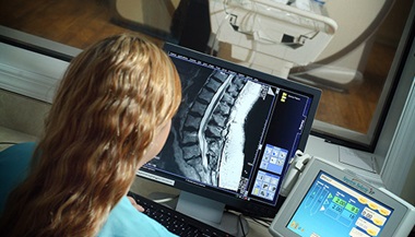
What injuries require a CT scan?
A CT scan, or computed tomography scan, sends radiation through the body. However, unlike a simple X-ray study, it offers a much higher level of detail, creating computerized, 360-degree views of the body’s structures.
CT scans are fast and detailed. They take longer than X-rays but are still fast (under one minute). This makes them ideal for emergency situations. “CT indications are often ordered for trauma, such as someone who was in an accident or fell, to rule out fracture, but they are also used for detailing complex bone anatomy prior to surgery” explains Fayad.
CT scans can spot:
- Blood clots
- Bone fractures, including subtle fractures not visible on X-ray
- Organ injuries
What are the differences between an MRI and CT scan?
A CT scan may be recommended if a patient can’t have an MRI. People with metal implants, pacemakers or other implanted devices shouldn’t have an MRI due to the powerful magnet inside the machine. CT scans create images of bones and soft tissues. However, CT scans aren’t as effective as MRIs at exposing subtle differences between the different types of tissue.
Talk to Your Doctor about Types of Imaging
You can play a more active role in your care by knowing the differences between a CT scan, MRI and X-ray. Don’t be afraid to ask your doctor why a particular type of imaging test has been recommended.
Often, your doctor will have consulted with a radiologist about what test you need.
“Radiologists are the doctor’s doctor,” explains Fayad. “We usually interface with other physicians, but patients are also welcome to talk to a radiologist directly if they have questions about an exam.”
The right imaging can lead to the right diagnosis, which is an essential part of effective treatment. It’s important to choose an imaging center that offers a full range of technologies, and experienced radiologists and technologists who are trained in specific body areas, diseases and imaging techniques.
Johns Hopkins Medicine offers a wide variety of imaging exams, including breast imaging, bone density (DEXA) scans, positron emission tomography (PET) scans, ultrasounds and wellness screenings.
Medically reviewed by Laura M. Fayad, MD

Talk to your doctor about types of imaging
You can play a more active role in your care by knowing the differences between a CT scan, MRI and X-ray. Don’t be afraid to ask your doctor why a particular type of imaging test has been recommended.
Often, your doctor will have consulted with a radiologist about what test you need.
“Radiologists are the doctor’s doctor,” explains Fayad. “We usually interface with other physicians, but patients are also welcome to talk to a radiologist directly if they have questions about an exam.”
The right imaging can lead to the right diagnosis, which is an essential part of effective treatment. It’s important to choose an imaging center that offers a full range of technologies, and experienced radiologists and technologists who are trained in specific body areas, diseases and imaging techniques.
Johns Hopkins Medicine offers a wide variety of imaging exams, including breast imaging, bone density (DEXA) scans, positron emission tomography (PET) scans, ultrasounds and wellness screenings.