Slipped Capital Femoral Epiphysis
What is the capital femoral epiphysis?
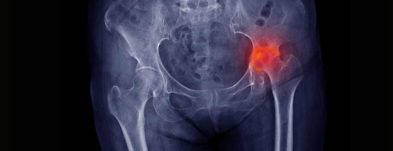
The femur is the long bone of the thigh. The end of the femur that connects with the hip consists of a “ball” (called the femoral head). The ball fits inside of a “cup” that is made up of the pelvic bones and is known as the acetabulum. During growth, the end of the head is known as the epiphysis and is connected to the rest of the femur by the growth plate.
What is slipped capital femoral epiphysis?
Slipped capital femoral epiphysis (SCFE) a disorder of adolescents in which the growth plate is damaged and the femoral head moves (“slips”) with respect to the rest of the femur. The head of the femur stays in the cup of the hip joint while the rest of the femur is shifted.
What causes slipped capital femoral epiphysis?
The exact cause of SCFE is not known. There are, however, many factors that are associated with this condition. These factors lead to weakening of the growth plate (also called the “physis”) which then causes the femoral head (ball of the femur) to slip off the neck of the femur. Obesity is a major risk factor. Certain endocrine disorders are risk factors for SCFE, such as hypothyroidism and osteodystrophy. There may be a genetic predisposition to this condition (it runs in families). Boys are more often affected than girls.
What are the signs and symptoms of slipped capital femoral epiphysis?
Patients may have pain in the groin, inner thigh, or knee. They may have stiffness and decreased ability to rotate the leg. In addition, there may be a change in their gait, or the way they walk, as they try to put as little weight as possible on the affected side. They may also walk with the leg rotated outward.
In most cases, onset of pain and/or limp is slow and gradual. However, symptoms can also occur suddenly and may be associated with a minor fall or trauma.
Slipped capital femoral epiphysis Diagnosis
The pediatric orthopaedic specialist will obtain a complete medical history and do a thorough physical exam. He/she will also order x-rays which will make the final diagnosis. In rare instances, an MRI may be needed if the x-rays are inconclusive.
Slipped capital femoral epipyhsis Treatment
The goal for treatment is to prevent the femoral head from slipping any further and to avoid complications. This is accomplished with surgery. A screw is inserted to connect the femoral head with the rest of the femur. If severe deformities are present, the surgeon may decide to realign the bones before placing the screw. The surgeon may also recommended placing a screw in the unaffected hip to prevent slippage before it occurs.
In some cases the slip is very unstable (may slip further rapidly) and so it is important that an orthopaedic surgeon sees the patient as soon as possible. In extreme cases, it is important to perform surgery on the day of diagnosis. However, most slips are stable and can wait 3-14 days for operative treatment.
During the time between diagnosis and surgery, it is important to have the child rest and avoid putting weight on the affected leg. For a period of time after surgery, the child will only be allowed to put limited weight on the leg and will require the assistance of crutches or a walker.
Are there complications from slipped capital femoral epiphysis or its treatment?
Unfortunately, there are several severe complications that can result from SCFE and its treatment. The first is “osteonecrosis.” “Osteo” means bone and “necrosis” means death. In osteonecrosis, the blood supply to the femoral head is damaged and the bone dies. This can lead to degenerative joint disease (osteoarthritis). The other complication is called “chondrolysis”. “Chondro” means cartilage and “lysis” means cutting apart. In this complication, the joint cartilage is damaged and leads to a painful and stiff joint.
What is the prognosis for patients with slipped capital femoral epiphysis?
How a patient will do depends on the severity and the presence of complications. Even without complications, there is still an increased risk of degenerative joint disease. Losing weight, however, is a major way in which an adolescent can reduce the risk of the later development of degenerative joint disease.