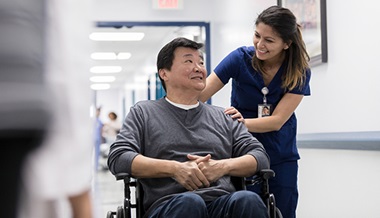
Patient Story
Pulmonary Valve Replacement: Michelle's Story
Patient Story Highlights
- Awais Ashfaq, MBBS saw that Michelle's pulmonary valve is degenerating.
- He reached out to Johns Hopkins all Children's Heart Institute to get a 3D model of her heart.
- The 3D model of her heart helped him gain a better understanding of the anatomy of the patient's heart.
Awais Ashfaq, MBBS, looks at the patient’s CT scans and MRI and realizes they aren’t enough. He needs more information. He needs to see her heart more clearly. He needs something tangible.
The patient’s pulmonary valve is degenerating. It was placed in her heart a decade ago when she was 1. It’s not uncommon for such a valve to need to be replaced, and often it can be done with a minimally invasive catheter procedure. But this case at Johns Hopkins All Children’s Hospital in St. Petersburg, Florida, is more complex.
The scans indicate the valve is crunched up. The right ventricle outflow track is deformed and a portion is incorporated in the sternum. Ashfaq, a cardiothoracic surgeon, knows open-heart surgery will be required to replace the valve, but he needs a better look at what he will encounter during surgery.
He needs a 3D model of her heart.
“Sometimes the scans do not reveal everything, especially the relationship of the left coronary artery, which usually sits right behind the pulmonary valve and can sometimes be stuck to that valve,” Ashfaq says. “That makes it hard to see everything with the naked eye.”
Starting the Process
Ashfaq emails Adam Verigan, an imaging informatics analyst who works closely with the Johns Hopkins All Children’s Heart Institute, to request a 3D print of the heart. Verigan returns a series of questions to Ashfaq to make sure he understands the specifics of the request.
“First, I have to make sure I understand what part of the patient’s heart the surgeon wants to focus on,” Verigan says. “You have to understand the software as well as the anatomy. It’s usually a complex anatomy when a 3D print is requested. Surgeons work with their hands, so in the end, by holding and seeing a model, and moving it around in their hands, they get size representation and other key information.”
Verigan sends a message to loop in Matt Lee, interim program manager and simulation engineer of the Center for Medical Simulation and Innovative Education at Johns Hopkins All Children’s.
Johns Hopkins All Children’s has collaborated with the USF Morsani College of Medicine for nearly 50 years. Once Verigan has the specifics of Ashfaq’s request and the appropriate images — including a magnetic resonance angiogram, a type of MRI that looks specifically at the blood vessels — from the Johns Hopkins All Children’s radiology team, he shares an anonymized version of them with the USF Radiology Department, which has a nationally known specialized 3D clinical applications team that can segment the image data and generate a 3D model file that translates into a 3D print.
“The most critical thing to this process is accuracy,” Lee says. “If it’s not accurate, it has no purpose at all. That’s why there are so many steps involved.”
New Technology
In 2020, Johns Hopkins All Children’s purchased a medical grade 3D printer, which can replicate complex cardiac properties, brain and spinal tumors, soft or elastic organs, craniofacial conditions and more. The printer offers medical professionals on-demand, ultra-realistic anatomical simulation models. The process works similarly to Ashfaq’s request for the 3D heart model.
“The use of 3D prints in preoperative planning is occurring more and more in the recent years,” he says. “It gives me a better idea of what I will find during the surgery.”
Lee operates the 3D printer in the Simulation Center at Johns Hopkins All Children’s. When he gets the 3D model file from the USF Radiology team, he shares it with Ashfaq for approval.
Generally, it takes about a week to get from request to approval of a 3D print project. Finally, it’s time to start the actual printing process.
It takes 15-21 hours to print a 3D model and another two to four hours for the cleaning process depending on how complex the model is and the material used.
In His Hands
Once it’s cleaned and ready, Lee hand delivers the printed replica to Ashfaq’s office, where the doctor confirms it meets his needs.
Ashfaq reviews the model and compares it with echocardiogram and CT scan images to gain a better understanding of the anatomy of the patient’s heart. He says in this case, the 3D model proves a big help.
“In situations where we need to do a lot of work inside the heart, or we need to create a pathway inside the heart, it can be invaluable,” he says.
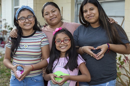
A Successful Outcome
The patient — Michelle — is back home now, living the life of an 11-year-old. She may not realize the teamwork and technology that enhanced her valve replacement. She’s focused on the here and now. She has a life of possibilities ahead.
“It’s a privilege to be a part of the process,” says Verigan, who coincidentally also was a heart patient at All Children’s as a child. “I’ve been a Johns Hopkins All Children’s Hospital employee for 14 years, but I’ve also been a patient in our heart program for 39 years. I would like to know that my doctor and surgeon have seen my anatomy using this technology and have had a chance to develop a surgical plan before surgery.”