Patient Story
Complex Spinal Reconstruction: Danica's Story
Complex Spinal Reconstruction | Danica’s Story

After a fall, 9-year-old Danica was left with metal rods, which had been implanted years before to stabilize her spine and had broken in the accident, floating dangerously close to her brain stem. Danica and her family traveled from Ohio to Johns Hopkins to receive life-altering, complex reconstruction surgery from Neurosurgical Spine Center Director Nicholas Theodore, M.D. Danica is now able to ride a bike for the first time ever.
Patient Story Highlights
The first sign of something wrong with Danica Snyder’s spine came at age 13 months, when she cried out in pain when turning her head a certain way during diaper changes. Then, while posing for a family portrait at 18 months old, Danica’s head kept falling to one side.

“Everyone said she had torticollis,” says Danica’s mother, Monica Kaye Snyder.
Torticollis, or “twisted neck” in Latin, which can occur due to positioning in the womb, can be corrected with stretching exercises at home. But physical therapy recommended by Danica’s pediatrician was not only not improving her condition, “she was getting worse,” says her mom. She then took Danica to a neurologist near their home in Uniontown, Ohio, who ordered a CT scan of her neck, which offered no clues to her problem. Then, she took her child to another neurologist, who ordered an MRI scan that revealed the culprit.
“We saw the Chiari malformation,” says Snyder, referring to a congenital defect in which brain tissue extends into the spinal canal.
Danica was referred to an Ohio hospital, where pediatric neurosurgeons were surprised to see excess bone along the right side of her cervical spine pulling her head to one side, signs of a rare condition called atlas assimilation. Indeed, vertebrae C2 and C3 were starting to fuse with the base of her skull, a condition no amount of physical therapy could fix. Complicating matters more, her brain stem and cerebellum had already started to drop into her spinal canal, compromising the flow of cerebral spinal fluid (CSF) surrounding and protecting Danica’s skull. The bones at the base of her skull needed to be opened up, a procedure called decompression, to allow for proper flow of CSF.
“There was no flow at all on the right side of her brain,” says Snyder.
Surgical Intervention
Surgeons successfully decompressed Danica, then age 2, but her crossing with Chiari malformation was far from over. Neurosurgeons scanning her six months later found further descent of brain tissue into her spinal canal and instability where the spinal cord meets the brain—the craniovertebral junction (CVJ). She needed to be decompressed again and have her spine stabilized with spinal instrumentation.
Danica underwent another decompression operation, but surgeons recommended she wait until she reached age 6 for fusion surgery. Seeing 3-D imaging of Danica’s Chiari, Snyder says: “I knew that wasn’t the answer. Part of the brain that had fallen was already dead.”
She found a neurosurgeon and spine surgeon who together would do Danica’s fusion surgery. After the surgeons stabilized the CVJ with metal rods and sewed in a cerebellar sling to keep her brain from falling again, they restricted her to a wheelchair and no physical activity for at least six months. Also during that time, she would have to wear a brace 24/7 for at least six months to help immobilize her spine. She ended up wearing it for a year.
“That was a very long journey for a young child to have no motor development activity,” says Snyder. “But we see now how that shaped Danica into who she is—a reader, musician, a functionally bright kid.”
But Danica also wanted to be like other kids, to play with them on the school playground when she was told she would have to sit on the sidelines. One day she said no, walked toward the playground and fell.
Indeed, Danica and her mom then heard clicking and popping sounds of metal on metal when Danica moved her head. She lost stability in her spine and, worse, the broken rods were endangering her brain. To further complicate matters, Danica’s last two surgeons had retired. Snyder tried to remain hopeful but knew there were few experts in the country who could manage a very complex patient like Danica.
“I didn’t know where else to go, where to get another opinion,” she says. “We were worried no one would take her case.”
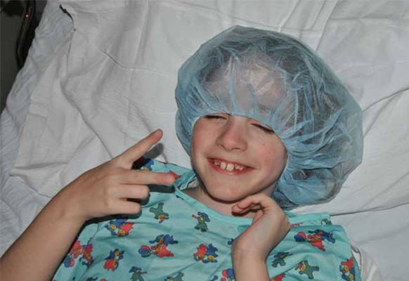
Danica prepares for surgery giving the peace sign and a big smile of confidence.
Technological Advancements Guide a Complicated Case
Then, she learned that one of those experts, neurosurgeon Nicholas Theodore, was moving from Arizona to Baltimore and The Johns Hopkins Hospital, which she knew well from working for many years in the Baltimore-Washington area. But he had just arrived and had not even settled into an office.
“He didn’t even have paper clips,” says Snyder. Nonetheless, Theodore’s longtime assistant, Julie Zeuch, said, “Send me Danica’s files.” Then Theodore, who had deep experience treating patients with motor vehicle-related traumatic spine injuries in Arizona, called. “He said, I’m honored and humbled to take this case. This is what I do,” recalls Snyder.
Indeed, Theodore had performed some 150 of these high-risk procedures. But as confident as he was, Theodore knew this revision spine surgery would be no walk in the park. He would be operating on the CVJ, the holy grail of spine surgery, what Theodore calls “high-priced real estate.” And this anatomy had been altered by broken rods and previous surgeries, obstructing his visual field.
“If you haven’t seen this before, from a surgical perspective, it’s kind of daunting,” says Theodore. “It’s like being in the middle of New York City and someone takes your map away and you don’t know where you are.”
To navigate this terrain, he took advantage of intraoperative imaging advances at Johns Hopkins, positioning Danica in an O-arm CT to obtain real-time 3-D images during the procedure.
“In this case, the technology was critical in determining the location of the broken rods, spinal canal and other critical structure all covered in semihealing bone,” says Theodore. “The imaging gave us immediate feedback where everything is.”
The imaging and Theodore’s surgical expertise were important in another regard too. In a previous surgery, the end of a spinal drain catheter had broken off and now threatened Danica’s brain stem. Using the intraoperative CT, Theodore knew exactly where it was and was able to neatly remove it instead of making a large opening in the dura—the membrane covering the spinal canal and brain—exposing the spinal cord and fishing around for it.
“We were able to make a very small opening, dissect the catheter away from the brain stem and take it out,” says Theodore.
To stabilize Danica’s severely unstable CVJ, which posed a risk of pinching her spinal cord and incurring severe neurologic deficits, Theodore meticulously removed and repaired the broken hardware in the six-hour operation. He also harvested a 3-inch piece of the girl’s top rib for additional support. This is natural bone, explains Theodore, which gives patients a higher rate of healing than cadaver bone.
“Also, the rib has a nice curve to it and fits perfectly between the base of the skull and the upper cervical spine,” says Theodore. “It’s as though God invented it as a piece to fill in this piece of the jigsaw puzzle.”
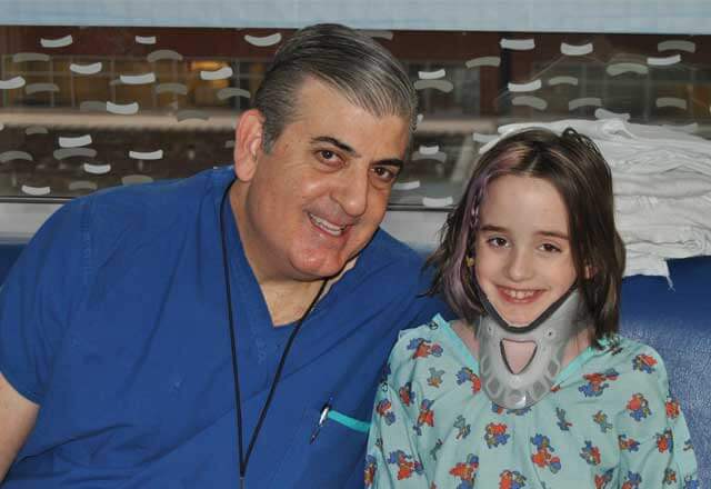
Danica and Dr. Theodore pose for a photo together in the Johns Hopkins Children's Center.
A Collaborative Win
Danica’s outcome?
“The pain she was having before is gone; she’s doing quite well,” says Theodore. “If she does end up having a solid fusion and everything heals perfectly, I think the future is bright for her. I think it is now. I’ve had pretty good luck in these revision operations.”
Theodore adds that the credit for such outcomes goes to the diverse skills and collaboration of Johns Hopkins’ surgical teams.
“This case underscores our different expertise and team approach history. It’s not one guy doing everything,” says Theodore. “This is one of those areas—complicated spinal problems—where we come together for the benefit of the patient. That type of collaboration is another reason I’m here.”
For Snyder, Danica’s recovery “was everything we had hoped for. She had zero pain after her surgery, and now she’s doing so well. We’ve had amazing support in the community and at school. She’s very loved.”
She attributes her outcome not only to Theodore’s surgical skills, but also to the way he interacted with Danica and infused her with confidence.
“The day before her surgery, Dr. Theodore came into Danica’s room and went right to her and spoke to her like there was no one else in the room. He asked her how she was feeling and talked about her operation, which gave her a lot of confidence going into the surgery,” says Snyder. “He made all the difference in restoring a childhood that she honestly never had.”
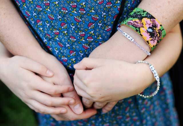
Danica and her mother wear matching bracelets signifying hope and trust.
Learn more about Chiari Malformation.
Danica's Neurosurgeon
The Chiari Malformation Center
The Johns Hopkins Chiari Malformation Center provides expert care to both children and adults with all types of Chiari malformation and related conditions like syringomyelia. Our doctors offer seamless treatment plans for patients of all ages, partnering with you from infancy into adulthood.