Selective Dorsal Rhizotomy (SDR)
What You Need to Know
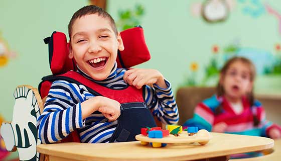
- Spasticity (muscle tightness) is common in children with cerebral palsy, and it can cause discomfort and difficulty during movement.
- Selective dorsal rhizotomy addresses spasticity by cutting select sensory nerve roots in the spine to stop the sending of abnormal signals that cause muscles to tighten.
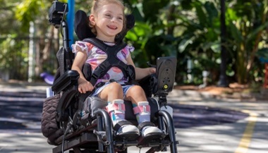
- Children as young as 3 can have SDR. Neurosurgeons, as part of a multispecialty team, evaluate many factors to determine if a child will benefit from the procedure.
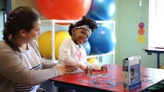
- Rehabilitation therapy is crucial to the success of SDR, and can last for several months.
What is selective dorsal rhizotomy?
Selective dorsal rhizotomy is a surgery performed in the lower spine that can help reduce leg spasticity (muscle tightness) in children with neurological disorders such as cerebral palsy.
During SDR, the neurosurgeon uses a microscope and electrophysiological monitoring to guide the cutting of abnormal sensory nerve rootlets that originate from the back (dorsal) side of the spinal cord. A specially trained neurologist assists the neurosurgery team by assessing and interpreting the monitoring data to help identify the dorsal nerve rootlets with the most abnormal responses. The neurosurgeon cuts the selected nerve rootlets. That’s why this type of rhizotomy is called selective, as opposed to a nonselective rhizotomy where monitoring is done only to preserve the motor and bowel or bladder roots.
This procedure is performed under general anesthesia.
Who performs selective dorsal rhizotomy?
Selective dorsal rhizotomy is performed by a pediatric neurosurgeon in close collaboration with a larger multispecialty team. The team includes pediatric neurologists and anesthesiologists, as well as specialists in orthopaedics, genetics, child development and child life. There are several specialized centers across the U.S. that have cerebral palsy programs with expertise in SDR.
Selective Dorsal Rhizotomy: FAQs with Dr. Shenandoah Robinson

Shenandoah Robinson, a Johns Hopkins Children’s Center neurosurgeon, discusses the opportunities for selective dorsal rhizotomy (SDR) in cerebral palsy patients.
Who can benefit from selective dorsal rhizotomy?
The child’s neurosurgeon, in collaboration with a multispecialty team, will be best able to determine if SDR is an effective treatment for their condition. SDR may be considered after treatments like physical therapy, botulinum toxin injections and medications do not provide adequate relief of the spasticity.
Neurosurgeons consider several factors when evaluating who is a candidate for SDR:
Which body parts are affected by spasticity: Children who have spasticity primarily in their legs usually benefit from SDR more than those with spasticity in their arms and legs. SDR is typically recommended for children with the type of cerebral palsy called spastic diplegia, which means muscle tightness affecting both legs. SDR is also effective for children with hemiplegia, when one leg is much more affected than the other.
Cognitive ability: Children with the cognitive ability to follow directions are good candidates for SDR given their ability to participate in extensive rehabilitation. Children with less ability to participate in a rehabilitation program may benefit less from SDR.
Level of physical function:
- Children who can walk or have the potential to walk with the aid of braces or walkers have the biggest potential to benefit from SDR. Children who experience severe muscle weakness are generally not SDR candidates as their level of function may worsen after the procedure.
- Some children with severe spasticity who can’t walk and have poor motor control can also benefit from SDR. In this case, the goal of the procedure is less to improve function and more to reduce discomfort, improve positioning and ease caregiving activities such as bathing. This approach is also called palliative SDR, and it sometimes involves cutting both the motor and sensory nerve roots in a specific area, not just the sensory ones.
SDR is not generally recommended for children with spastic diplegia and significant muscle weakness. These children often rely on spasticity to compensate for the lack of muscle strength in affected areas. SDR is also not generally recommended for children with severe dystonia (variable muscle tone).
If SDR is not recommended, the care team might suggest other options, including:
- Intrathecal baclofen pump, which is a pump surgically added into the abdomen that administers baclofen (muscle relaxant medication) via a tiny catheter directly into the spinal fluid.
- Orthopaedic procedures. Some children who undergo SDR will also benefit from orthopaedic procedures such as tendon lengthening at some point after the SDR. In rare cases, SDR may be combined with an orthopaedic procedure during the same surgical visit.
- Children with some types of dystonia may benefit from deep brain stimulation or intrathecal baclofen pump therapy.
When is the best time to have SDR?
Because children will need to participate in intensive therapy after the SDR, those who can cooperate with the therapy benefit most from the procedure. Very young children, such as those younger than 3, generally do not yet have the cognitive ability to be motivated to do rehabilitation and to follow instructions.
Having SDR before starting grade school, which typically starts at age 5, can help children increase their independence and keep up with their peers better. They can focus less on moving and more on learning and making friends.
However, older children, adolescents and adults who meet certain criteria may also benefit from SDR.
Selective Dorsal Rhizotomy for Adults
While less common, adults can also have SDR. Although results have been mixed, some studies show that people who have SDR as adults can experience improvement in walking, sitting, standing and balance, as well as increased endurance and ability to be active. Side effects include numbness, loss of sensation and a greater risk of chronic pain. Adults may also greatly benefit from intrathecal baclofen (ITB) therapy.
People who have lived with spasticity for a long time may experience signs of early aging. The signs include premature muscle weakness, pain and decreased mobility. SDR or ITB can help slow this process and delay these symptoms in adults.
After SDR, the patient, whether child or adult, needs to relearn how to control muscles and move without spasticity. This process might be more challenging for adults, who have lived with spasticity longer than kids.
Types of Selective Dorsal Rhizotomy
SDR is commonly done through a surgical approach called single-level laminectomy SDR. Typically, there are multiple muscles affected by spasticity, and the misfiring nerves originate near the same level of the spine. With this approach, the surgeon removes a small piece of bone from the back of the vertebra through a procedure called laminectomy. Then under the microscope, multiple sensory nerve roots are tested and selected ones are cut.
In the past, multilevel laminectomy SDR was used, in which bone was removed from several vertebrae of the spine. However, this approach has been largely phased out due to availability of a less-invasive, single-level procedure.
Selective dorsal rhizotomy is typically biliteral, meaning that nerve roots are cut on both sides of the spine. This approach is also used for children with hemiplegia, who have one leg affected more than the other. SDR is rarely unilateral, meaning it is rarely performed only on one side of the spine.
Spastic Cerebral Palsy Surgical and Rehabilitation Treatment | Journee’s Story
Journee, age 4, was born with cerebral palsy. She and her mother traveled from Boston to meet pediatric orthopaedic surgeon Ranjit Varghese and neurosurgeon Shenandoah Robinson. Journee's surgeons developed a treatment plan with the rehabilitation team at the Kennedy Krieger Institute to get her standing and walking for the first time.
Preparing for Selective Dorsal Rhizotomy
The care team may conduct a variety of tests and assessments to confirm that the child is a good candidate for the procedure, and to help plan the surgery. These tests and assessments may include:
A comprehensive evaluation by a multispecialty team, which includes specialists in pediatric neurosurgery, neurology, developmental pediatrics, orthopaedics, genetics, physical therapy and child life. This evaluation involves a physical exam and a review of medical history to determine the level of disability and response to previous treatments.
An MRI scan of the brain to assess the degree of scarring in the white matter (area of the brain that transmits information to other parts of the nervous system) and look for other changes in the brain. The results of the scan are also used to estimate how much a child might expect to improve after SDR.
Imaging of the spine to account for any anatomical differences during surgery.
What happens during SDR surgery?
To perform the SDR procedure, a neurosurgeon makes a cut in the lower part of the spine to remove a small section of bone and expose the nerve roots that run below the spinal cord. Typically, the bone is removed from one or two levels of the spine.
Next, the neurosurgeon identifies the sensory (dorsal) nerve roots (the ones carrying sensation signals) and separates them from the motor (ventral) nerve roots (the ones carrying movement signals). Spasticity results from abnormal signals sent through sensory nerves, so they are the focus of the surgery while the motor nerves, as well as nerves to the bladder and bowel, are protected. The surgeon also needs to determine which sensory nerves carry abnormal signals.
To accomplish this, the neurosurgeon uses a special surgical microscope to see the tissues within the spine and performs electrophysiological testing (electromyography) to stimulate each nerve root. A neurologist monitors how the child’s leg muscles respond after each nerve rootlet is tested, notes the abnormal responses, and provides feedback to the neurosurgeon. The surgical team follows this process on each side of the spine.
The selected sensory nerve roots are divided to expose the strands of nerve fiber within, called rootlets. Each rootlet is then tested with electromyography to confirm which one to cut. Approximately 15–25 rootlets are tested on each side.
After the most abnormal nerve roots have been cut, the surgical team carefully closes the wound layer by layer.
The entire SDR procedure takes about four hours to complete.
Recovery After SDR Surgery
Hospital Stay
Following surgery, the child will need to stay in the hospital to be monitored for a few days. The child will need to lie flat for a day or longer to protect the surgical area. Medications are available if the child needs help lying calmly. The child will also be given intravenous (IV) pain medication, as the nerve stimulation during the surgery can be uncomfortable for 24–36 hours after the surgery. The surgical team works in partnership with the anesthesiology team to reduce any discomfort and anxiety.
What to Expect
Spasticity will be noticeably reduced after the surgery because the operation cuts the nerves that carry abnormal signals resulting in muscle tightness. After spasticity is reduced, any underlying muscle weakness may become more obvious. The child often feels weaker at first and will need to gradually gain muscle strength.
In addition, the child will need to relearn how to walk with an improved gait without the effects of spasticity. Several months of inpatient and/or outpatient rehabilitation can help with both of these challenges.
Rehabilitation Therapy
At discharge from inpatient rehabilitation, the child and family will receive instructions for prescribed medication and guidelines for resuming physical activity and returning to school. The care team will help schedule physical and occupational therapy sessions that typically take place several times a week for three to six months. The exact schedule will depend on the child’s condition and needs. Some children need inpatient therapy followed by extensive outpatient therapy. Children may also be introduced to new equipment, such as different types of braces.
Selective Dorsal Rhizotomy for Spastic Diplegia: Landon’s Story

Outlook for Children After SDR
The long-term improvement from SDR will depend on the child’s condition before the surgery and how closely they follow the recommended rehabilitation protocol. In general, kids will go up one device level, meaning if they needed a walker before the surgery, then they may be able to walk with canes after surgery.
For children with spasticity primarily in the legs, SDR with physical and occupational therapy can lead to:
- Decrease in pain from spasticity
- Improved mobility and walking
- Increased stamina
- Improved balance
- Better standing and sitting posture
For children with spasticity in both arms and legs, SDR with physical therapy can lead to increased ability in the child to comfortably sit up.
For children with both arms and legs affected by spasticity, or those with a combination of spasticity and dystonia, the intrathecal baclofen pump therapy often provides greater relief, improved comfort and better functional improvement than a palliative SDR.
Selective Dorsal Rhizotomy Risks and Complications
Selective dorsal rhizotomy carries low risk of infection and bleeding. Because the surgery is performed on the spine, there is a risk of a cerebrospinal fluid leak. This is why the child needs to lie flat after surgery.
The cutting of the sensory nerve roots will not lead to paralysis, but it can rarely (less than 1%) result in:
- Increased sensitivity to touch stimulation (hyperesthesia)
- Worsened weakness that impairs ability to walk
- Issues of the back, including pain, spine curvature and slipped vertebrae (spondylolisthesis) after a multilevel laminectomy SDR.
Because of the careful monitoring during the surgery, and how the nerves to the bladder and bowel are protected during the surgery, the risk to bladder and bowel function leading to incontinence is very low.