Understanding Mammogram Results
A mammogram report is a summary of findings from your screening or diagnostic mammogram. Your doctor may explain the results to you, but if you use an online patient portal such as MyChart, you should also receive a copy of the report in the mail or in your patient portal.
While it is always best to contact your doctor to clarify the findings of the report, here are answers to some commonly asked questions:
Who makes the mammogram report?
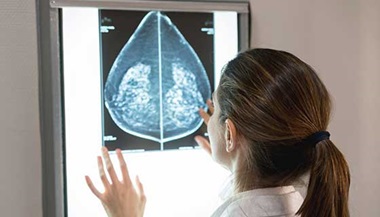
After the mammogram is completed by the mammogram technologist, the images are read and interpreted by a breast-imaging radiologist, who prepares the mammography report and sends it to your doctor and your online portal. You may be able to see your results before you’ve heard from your doctor, but it’s a good idea to wait for your doctor’s review so you can fully understand them.
What is included in a mammogram report?
A typical mammogram report contains the following:
- Relevant patient history
- Reason for the exam
- Whether previous mammograms were used for comparison
- Description of the findings
- BI-RADS category score (see below)
- Radiologist’s recommendations
What is the BI-RADS category on my mammogram report?
The Breast Imaging-Reporting and Data System (BI-RADS) is a standardized set of definitions required by the federal government and used by radiologists to describe mammogram findings. The scores are used to help doctors classify any notable details of the images and decide next steps.
For breast imaging, the BI-RADS scores range from 0 to 6:
- BI-RADS Category 0: Incomplete: Additional imaging evaluation or comparison to earlier mammograms is needed.
- BI-RADS Category 1: Negative: No abnormal areas found.
- BI-RADS Category 2: Benign: No cancerous findings, but showing some areas of abnormality such as calcified areas, masses, cysts or scar tissue that are not breast disease.
- BI-RADS Category 3: Probably benign finding: There is less than a 2% chance of being cancer, but it warrants a prompt follow-up.
- BI-RADS Category 4: Suspicious abnormality: Biopsy should be considered.
- 4A: 2% to 10% likelihood of being cancer
- 4B: 10% to 50% likelihood of being cancer
- 4C: 50% to just under 95% likelihood of being cancer
- BI-RADS Category 5: Highly suggestive of malignancy (95% or greater likelihood of being cancer): Biopsy strongly recommended.
- BI-RADS Category 6: Known, biopsy-proven malignancy. Mammograms can test the area of cancer in the breast to see how it is responding to treatment.
For mammographic screening, BI-RADS categories of 0, 1 or 2 are given. For diagnostic imaging, BI-RADS categories 1–6 can be given.
Treatment Did you know?
How do I know if my mammogram results show something abnormal?
If your mammogram report shows BI-RADS Category 0, the study is considered inconclusive, and you need to come back from more imaging.
If your report shows BI-RADS Category 1 or 2, your mammogram is completely normal or has benign findings that do not require additional imaging or follow-up. If you do not notice any breast changes, nothing else needs to happen until your next scheduled screening mammogram.
BI-RADS Category 3 indicates that there is something in the breast that is considered probably benign, and a six-month follow-up is recommended.
BI-RADS Categories 4 and 5 indicate that there is an abnormality that requires a biopsy. BI-RADS Category 6 is reserved for breast cancer that has already been diagnosed with biopsy.
What are the most common reasons for being called back after a mammogram?
If you have a BIRADS 0 on your screening mammogram, you will need to return for a callback mammogram and/or an ultrasound exam. If this happens, do not panic. About 1 in 10 mammograms result in a callback. In about 90% of these cases, further testing reveals nothing serious. Callbacks are more likely after your first mammogram because there are no previous images to compare it with, which prevents the radiologist from identifying anything new and different. Getting more screening mammograms over time will help radiologists note the normal appearance of the breasts and make it easier to notice any changes.
Here are some of the terms used to define abnormal mammogram results that may lead to being called back for more testing:
Asymmetry:
An asymmetry is when there is an area of breast tissue that looks denser than other areas of breast tissue.
Should I worry about asymmetry in a mammogram?
A callback for an asymmetry is not cause for alarm. It means more imaging is needed for a better understanding.
After a callback for an asymmetry, a breast-imaging radiologist will obtain additional mammogram views and often results from a targeted ultrasound exam. Frequently, asymmetries represent normal breast tissue. But if the asymmetry has any suspicious features, the radiologist will recommend a follow-up exam or a biopsy.
Breast Calcifications
Calcifications are calcium deposits in the breast tissue. They are very common, and the great majority do not indicate cancer. Some early types of breast cancer may present as calcifications. If a radiologist sees calcifications that are not definitely benign, they may call you back for additional mammogram images that are designed to view calcifications better. After these additional images, the radiologist will decide whether a follow-up exam or a biopsy is necessary.
Architectural Distortion
Architectural distortion refers to a distorted or irregular appearance among the different tissue patterns in the breast (fat, blood vessels, ligaments, glands and fibrous tissue) without evidence of a mass or an abnormal dense area. Architectural distortion may be a sign of benign (noncancerous) scar tissue or cancer.
Breast Cyst
A breast cyst is a pocket of fluid within the breast tissue. On a mammogram, a cyst usually looks like a smooth, round mass. A radiologists cannot definitely differentiate a breast cyst from a solid mass on a mammogram. For that reason, they may recommend an ultrasound exam, which allows them to confidently distinguish a benign cyst from a solid mass, which may require a biopsy.
Learn more about breast cysts and other benign breast lumps.
Breast Mass
A breast mass usually appears as a white, dense spot on a mammogram. A mass is not always cancerous, and radiologists know how to tell a suspicious mass by examining its shape, edges and location. An ultrasound is almost always used to further evaluate the mass. Not all breast cancers, especially in early stages, appear as a mass. Further testing such as a biopsy can help confirm the type of cells that make up the mass.
Breast Density
If you have been told you have dense breasts, you’re not alone. In fact, 45% of women have dense breasts. Inside the breast is a combination of fat and glandular and fibrous tissue. Dense breasts have a lot of glandular tissue and less fat. This fibroglandular tissue appears white on the mammogram and makes abnormal findings, including cancer, harder to see.
Breast imaging reports inform patients about their individual breast density status. This is part of new federal guidelines that serve to increase reporting transparency between providers and patients.
The BI-RADS assessment describes four categories of breast density that you may see in your mammogram results:
- Category A: The breasts are almost entirely fatty (it is easier to see abnormal findings in fat than in fibroglandular tissue).
- Category B: There are scattered areas of fibroglandular density, meaning there are dense areas within the breast.
- Category C: The breasts are heterogeneously dense, which means they are dense throughout — not just in a few areas. This may obscure small masses.
- Category D: The breasts are extremely dense, which lowers the sensitivity of mammography.
Follow-Up Tests After a Screening Mammogram
If your mammogram results include one or more of the above findings, the radiologist may recommend additional testing. These tests can provide a different or better view of the suspicious or unclear area, in order to better determine whether the abnormality found on the screening mammogram is benign or related to cancer. Additional tests may include:
- An additional (diagnostic) mammogram
- Breast ultrasound
- Breast MRI
- Breast biopsy
Studies have shown that radiologists who specialize in reading only breast imaging detect more cancers and more early-stage cancers and have lower callback rates than general radiologists who read images of all types of body parts.
What if I have breast cancer?
If a biopsy reveals the presence of breast cancer, you will be referred to a breast surgeon to discuss next steps in your care.
The biopsy will provide the doctor with information about cancer type.
The thought of getting a breast cancer diagnosis is frightening, but it’s not a reason to avoid screening. Having regular mammograms means that if you do develop breast cancer, it is more likely to be discovered at an early stage, which can mean a much better chance of good treatment outcomes.
What should I do if I notice abnormal changes or symptoms after my mammogram comes back normal?
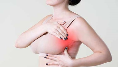
Regardless of when you had your last mammogram, contact your doctor any time you notice anything abnormal in a breast or nipple such as a lump, swelling, distorted shape or unusual discharge. Do not wait until your next mammogram unless your doctor says it is OK. Breast pain is seldom caused by cancer, but if it persists, contact your doctor.
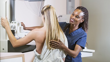
Schedule Your Mammogram

We have mammograms and other imaging available in the following locations:
All Johns Hopkins Medical Imaging sites exclusively offer 3D mammograms.