Conditions We Treat: Complicated Monochorionic Twins
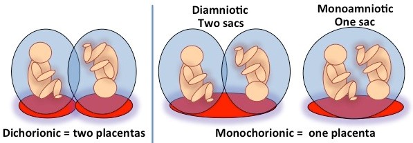
During normal pregnancy, the placenta is the source of nutrient exchange between the mother and her fetus. The blood vessels in the umbilical cord of the baby are responsible for linking the circulation of the baby to the circulation of the placenta to allow this nutrient exchange. When there is more than one fetus, such as a twin or triplet pregnancy, two types of placentation are possible. Most common is dichorionic placentation, where each fetus has its own placenta. When both fetuses share one placenta, this is called a monochorionic placenta.
Common Characteristics of Monochorionic Placentas
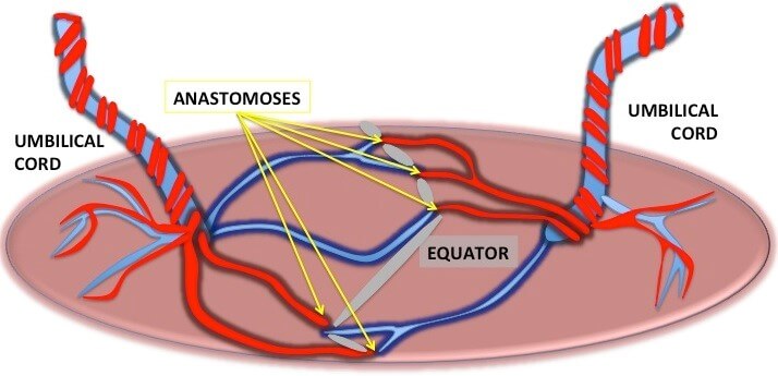
All monochorionic placentas share some common characteristics. The umbilical cord of each fetus inserts into the surface of the placenta, and blood vessels that run on the surface of the placenta between these cord insertions. These blood vessel connections can be between an artery of one baby and the vein of the other baby (AV anastomosis), between an artery from each baby (AA anastomosis) and between two veins from each baby (VV anastomosis).
Because blood pressure is higher in arteries, AV anastomoses allow blood or volume exchange from one baby to the other, a process called unidirectional shunting. The direction of blood flow in AA and VV anastomoses depends on which baby happens to have the higher pressure in these vessels and therefore can fluctuate between sides, a process called bidirectional shunting. Sometimes, shunting between these anastomoses can switch back and forth from one heartbeat to the next.
Each baby has a share of the placenta that is available for its nutrient delivery. The area of the placenta where the blood vessels of both babies meet is called the vascular equator, because it is the natural dividing lines between the portions of the placenta that belong predominantly to one baby or the other. The position of the vascular equator determines the placental share of each baby.
In uncomplicated monochorionic twin gestations, the blood exchange between both babies is equal, and the placental mass is equally shared. But one-third of monochorionic twin pregnancies are at risk for complications when sharing between both babies is not equal.
Complications of a Monochorionic Placenta
Complications that can arise in monochorionic pregnancies are due to unequal sharing of blood, blood volume, placental nutrients or a combination of these. Essentially the existence of anastomoses between twins links the well-being of each twin to the other. These connections are believed to be responsible for the higher rate of developmental delay found in complicated monochorionic twins.
Complications such as TTTS, TAPS and SIUGR may present as distinct or mixed clinical pictures. Because these situations require different therapies, accurate prenatal diagnosis and expert fetal and maternal surveillance is critical to maximize the chances for a favorable outcome.
Diagnosing Complicated Monochorionic Twin Pregnancy

The first and most critical step in identifying risks for complications in multiple pregnancies is to determine whether the placenta is monochorionic. This is best done in the first 12 weeks of pregnancy— the first trimester—by a prenatal ultrasound. The finding that identifies a monochorionic placenta with a high level of certainty is the so-called lambda sign, an ultrasound finding in those with monochorionic placentas.
Once it has been determined that the placenta is monochorionic, detection of complications requires close attention to signs of growth discordance, volume discordance or discordant blood counts between fetuses. In addition, a detailed and sometimes repetitive assessment of the anatomy of both fetuses is required. This is done by advanced ultrasound techniques using high-resolution scanning, Doppler techniques and three-dimensional imaging. Because the conditions can evolve, ongoing surveillance is required to detect deviations of the clinical course that may require specific therapy.
Management Options for Complicated Monochorionic Twin Pregnancy
The management expertise that is required for complicated monochorionic twins depends on the specific conditions.
- Fetoscopic laser occlusion of placental anastomoses offers the best outcome for pregnancies complicated by TTTS or TAPS. Developing the equatorial dichorionization, Dr. Baschat has been able to achieve survival rates over 95 percent coupled with minimal recurrence risk for pregnancies complicated by TTTS.
- Cord occlusion offers the best chance for one baby. This is a management option for pregnancies complicated by severe SIUGR, where it is deemed unlikely that the growth-restricted baby will survive, or where discordant fetal anomalies or TRAP sequence are present. Following cord coagulation, the normal co-twin has an over 90 percent chance for an uncomplicated pregnancy course.
- Fetal surveillance is complex, because multiple aspects of the condition in each fetus can change. Dr. Baschat has long-standing experience applying his integrated surveillance approach. This surveillance method incorporates a comprehensive list of maternal and fetal surveillance parameters to determine how often patients need to be seen and when intervention of delivery is advisable.
The decision of which treatment option is the most appropriate depends on a detailed assessment by high-resolution 2- and 3-D ultrasound, fetal Doppler flow studies, and sometimes additional magnetic resonance imaging. The Johns Hopkins Center for Fetal Therapy combines world-class expertise in all required imaging and therapeutic modalities in one location.