Our Research Facilities

Our Core Facilities and Shared Instruments

Jeol NMRThe NMR facility is located in Hunterian SB2 and accessible to all researchers at JHMI (training required and billed quarterly)

Pharmacology NMR Facility

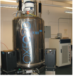
To facilitate research involving small molecules, the Department manages a Jeol ECZ500R, 500 MHz NMR spectrometer equipped with a Royal HFX probe, a 24-position autosampler, and a VT system (−150 to +150 °C). The Royal HFX probe is capable of direct ¹H and ¹⁹F detection either separately or simultaneously (single tune versus HF-dual tune). While locked on ²H it can also detect ¹³C and ³¹P and all nuclei between ³¹P to ¹⁵N (includes e.g. ¹¹B, ¹⁷O, ²⁹Si, ³³S, etc.).  The automation is configured for a convenient queue up of common 1D (¹H, ¹⁹F, ¹³C, ³¹P, DEPT, ¹H with solvent suppression) and 2D (COSY, TOCSY, HSQC, HMBC, and NOESY) experiments. The facility is open to members of the JHU community including JHU technology ventures. As this spectrometer was acquired through an NIH award all research publications and grant applications pertinent to the use of this NMR must be acknowledged as follows. "NMR data were acquired using the JHU-Pharmacology JEOL JNM-ECZL500R spectrometer supported by NIH Instrumentation Program award S10OD034217."

Shridhar Bhat, Ph.D.
Facility Manager
Pharmacology and Molecular Sciences
Johns Hopkins University School of Medicine
725 North Wolfe Street / Hunterian 301
Email: [email protected]
Phone: 410-502-4803

Cryo-EM FacilityThe Cryo-EM is located in Hunterian SB3 and accessible to Pharmacology researchers only (training required)

Cryo-EM Facility

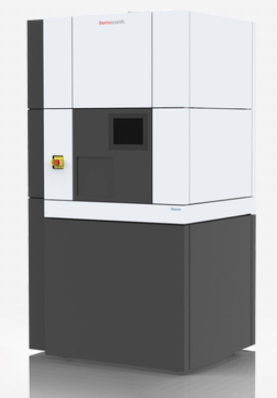
Pursuant to a generous evaluation agreement with Thermo Scientific, Pharmacology maintains a Tundra Cryo-EM to advance structural analyses of biomolecules bound to small-molecules using cryo-electron microscopy. Tundra operates at an accelerating electron voltage of 100 keV and can generate high-contrast 3D images of biomolecules and their complexes down to 2.6 Å at a medium throughput. Tundra comes with a cryo-loading station (CLS) that makes sample loading/unloading quick and easy coupled with simplified data collection. Thanks to AI-enabled smart single-particle analysis (SPA) data acquisition EPU-2 software Tundra can analyze intermediate results, provide instant feedback, and steer data collection on the fly. Thus, without extensive training new users can quickly optimize sample conditions. If a higher resolution structure is required, prescreened samples can be analyzed on more powerful Cryo-EM systems including those at the JHU Beckman Center (Krios, 300 keV; Glacios, 200 keV; Aquilos-2 cryo-FIB/SEM). Users should acknowledge the generosity of Thermo Scientific in publications reporting data based on the Pharmacology Tundra Cryo-EM.

Data Analyses
To perform single-particle analyses (SPA) and 3D reconstruction of the 2D projection images acquired on Tundra, Pharmacology has set up a server (comprising—4x Nvidia RTX A500 GPUs, 2x Intel Xeon Gold, 18-core CPUs with 1TB RAM and 2TB SS-hard drive, and 120 TB storage drive) and two front end workstations (Intel i9 x32 cores, Nvidia GeForce RTX 4090, 32 GB RAM; connecting to the server via an OM4 fiber optic network). These Linux workstations can run (locally or via the server) cryoSPARC, Relion and Scipion—the three prevailing SPA and 3D reconstruction programs.

Cryo-EM Sample Preparation
In order to prep the cryo-EM grids for sample deposition, a Pelco easiGlow plasma glow discharge apparatus is set up in Physiology-311. The samples can then be blotted onto plasma cleaned grids and vitrified using a Thermo Scientific Vitrobot (model: Mark IV) installed in the same room.

Shridhar Bhat, Ph.D.
Facility Manager
Pharmacology and Molecular Sciences
Johns Hopkins University School of Medicine
725 North Wolfe Street / Hunterian 301
Email[email protected]
Phone410-502-4803

Mass Spectrometers

Pharmacology has two mass spectrometers, an Agilent 6546 LC-QTOF, and a Bruker timsTOF flex. Access is restricted to trained researchers in the Department of Pharmacology.

Agilent 6546 LC-QTOFThe Agilent 6546 LC-QTPF is located in Physiology 318 and accessible to Pharmacology researchers only (training required)

Agilent 6546 LC-QTOF

Pursuant to a generous Equipment Placement Agreement between Agilent Technologies, Inc. and JHU-Pharmacology, Agilent installed a 6546 LC-QTOF system in March 2024. This LC-MS features high sensitivity, wide dynamic range, high resolution, and isotope fidelity, and accuracy of Q-RAI (Quadrupole Resolved All Ions). It is a workhorse instrument for small-molecule characterization and qualitative/quantitative metabolomics, oligonucleotides and proteomes. This LC-MS system consists of 1290 Infinity II binary pumps, temperature controlled multisampler and multicolumn compartment, diode array detector, dual AJS (Agilent Jet Stream) and ESI ion source, and 6546 QTOF (acquisition rate: 50 spectra/s, accuracy: <0.8 ppm RMS, sensitivity (S/N): 500:1 RMS, range (m/z): 20–3200).
Users should acknowledge the generosity of Agilent in publications reporting data from this LC-QTOF.

Data Analyses
A Windows workstation (AMD Ryzen 9 x16 cores, 32 GB RAM, Radeon RX 580 24GB GPU) is set up in Physiology-313 to process data acquired on the Agilent LC-QTOF and it has all the relevant software from Agilent to analyze small-molecule HRMS, metabolomics, oligonucleotides, and targeted proteomics LC-MS data.

Shridhar Bhat, Ph.D.
Facility Manager
Pharmacology and Molecular Sciences
Johns Hopkins University School of Medicine
725 North Wolfe Street / Hunterian 301
Email[email protected]
Phone410-502-4803

Bruker timsTof fleXThe Bruker timsTOF fleX is located in Biophysics B11 and accessible to Pharmacology researchers only (training required)

Bruker timsTOF fleX

Bruker timsTOF fleX (trapped ion mobility separation—Time Of Flight with dual source—MALDI and ESI) is an avant-garde mass spectrometer for PASEF (Parallel Accumulation–Serial Fragmentation) empowered 4D-Omics and it provides the capability to do high spatial resolution and high speed MALDI-MS as well as MALDI guided SpatialOMx® on tissue sections and high sensitivity ESI-MS. The timsTOF fleX is a one-stop mass spectrometer to analyze all kinds of small-molecules and biomolecules, including small-molecule metabolites, lipids, glycans, nucleotides, and proteins. Highly advantageous ion mobility parameter K (measured based on Collisional Cross Section [CCS] of the detected ion) is an integral element in timsTOF dataset and hence, in addition to mass and charge, very challenging isobaric interferences can be resolved and isomers can be distinguished and characterized fully.

Specifications of timsTOF—Acquisition rate: >100 Hz (in PASEF mode), Sensitivity: 50 fg/µL <15% RSD, Mass Accuracy: 800 ppb RMS, Mass Range: 20–40,000 m/z.

Introducing Samples into timsTOF
MALDI-MS measurements can be performed with samples spotted on MTP AnchorChip 384 which in turn is mounted on an MTP target frame (both are provided by the Facility).

The ESI-MS samples on the other hand are introduced using a Thermo Scientific EASY-nLC 1200 nanoflow LC system coupled to timsTOF via the CaptiveSpray ion source.

Shridhar Bhat, Ph.D.
Facility Manager
Pharmacology and Molecular Sciences
Johns Hopkins University School of Medicine
725 North Wolfe Street / Hunterian 301
Email: [email protected]
Phone: 410-502-4803

Pharmacology Shared Instruments

LM10 MicrofluidizerThe LEX-48 Parallel Bioreactor System is located in WBSB 312 and accessible to Pharmacology researchers only (training required)

Lex-48 Parallel Bioreactor System

To accelerate protein production (up to 40 L culture), Pharmacology maintains an LEX-48 Bioreactor from Harbinger Technology & Engineering. This is equipped with temperature control (heater/chiller circulator), climate control, and mixing is performed with direct aeration, leading to 20–50% higher protein yields.


LM10 Microfluidizer 2The LM10 Microfluidizer is located in WBSB 312 and accessible to Pharmacology researchers only (training required)

LM10 Microfluidizer

LM10 Microfluidizer is a pneumatically driven cell-lysing system for bacteria, yeast, insect, and mammalian cells (volume: 30–500 mL, microfluidizer pressure: up to 23,000 psi).


Beckman-Coulter Avanti J-E CentrifugeThe Beckman-Couter Avanti J-E Centrifuge is located in WBSB 312 and accessible to Pharmacology researchers only (training required)

Beckman-Couter Avanti J-E Centrifuge

A high-speed centrifuge (Avanti J-E with two rotors—JA-10 [typical use: cell harvesting, up to 3 L per run] and JA-20 [typical use: clarifying cell-lysates, up to 400 mL per run]).


Li-COR Odyssey CLx-2760 ImagerThe Li-COR Odyssey CLx-2760 Imager is located in WBSB 312 and accessible to Pharmacology researchers only (training required)

Li-Cor Odyssey CLx-2760 Imager

The Department members have access to this Li-COR imager to assist with visualization of fluorescent gels and immunoblots. The Odyssey CLX-2760 has two channels (700 and 800 nm) and thus it is capable of multiplexing.


BioRad ChemiDoc MP Imaging SystemThe BioRad ChemiDoc MP Imaging System is located in WBSB 312 and accessible to Pharmacology researchers only (training required)

BioRad ChemiDoc MP Imaging System

ChemiDoc MP Imaging System is an all-in-one system for imaging and analyzing gels and blots by detecting multiplex fluorescence (RGB, far red, near infrared, and StarBright dyes), chemiluminescence, and a wide range of stains (Coomassie, Silver, Ethidium bromide, SYBR, etc.). The most appealing feature of ChemiDoc is the ability to perform stain-free imaging of gels and blots—ChemiDoc achieves this by performing UV irradiation on-the-fly (that results in covalent bonding of tryptophan residues with a trihalo compound embedded in the gel/blot) and detecting the enhanced natural protein fluorescence—that enables total protein normalization (TPN)—an accepted and better alternative to using housekeeping protein as the loading control.


BioRad ChemiDoc MP Imaging SystemThe BioTek Synergy Neo2 Plate Reader is located in WBSB 312 and accessible to Pharmacology researchers only (training required)

BioTek Synergy Neo2 Plate Reader

Synergy Neo2 is a multimode plate reader equipped with BioStack plate stacker/loader (up to 50, 24- to 1536-well plates) that can record UV-Vis absorbance, fluorescence intensity, fluorescence polarization, FRET, TR-FRET, and BRET; and luminescence.