Research Story Tip: Scientists Create Map of Gatekeeper That Could Help Brain Cells Survive Stroke
11/10/2020
Researchers at Johns Hopkins Medicine have revealed the structure of a gatekeeping protein that could one day impact the treatment of conditions such as heart attack and stroke. Understanding the structure of the protein — known as the proton-activated chloride channel (PAC) — helps researchers develop ways to reduce permanent damage caused by conditions associated with acidosis, a condition marked by increased acidity in the blood.
A report of the study, was published Nov. 4, 2020 in Nature.
“Knowing the structure of the PAC helps us understand how this acid-sensing protein works in different pH [the measure of acidity or basicity of a solution] and gives us potential ways to manipulate it for better medical outcomes,” says Zhaozhu Qiu, Ph.D., assistant professor of physiology at the Johns Hopkins University School of Medicine and co-corresponding author of the study.
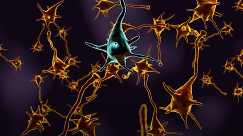
The PAC protein is activated when the environment around the cell becomes acidic. Typically, acidity in the body is held at normal levels through constant blood flow. However, when the circulatory system is disrupted by heart disease, stroke or some cancers, the area quickly becomes acidic as cellular waste builds up.
The Johns Hopkins Medicine-led research team first reported the gene sequences encoding this new “acid sensor” in Science last year. In the study, the researchers showed that stroke can over-activate PAC and kill brain cells in mice.
For the current study, Qiu’s team worked with collaborators, led by Wei Lü, Ph.D., assistant professor of structural biology at the Van Andel Institute and co-corresponding author of the study, to collect two types of images of the PAC. One was taken in the protein’s relaxed state — at a cell’s normal biological acidity level — and the other in its active state, when the environment is highly acidic.
To create an accurate picture, they used the specialized, high-powered cryo-electron microscope that supercools molecules so they form precise, easily imaged structures at sizes nearly to the atomic level. The images showed that the PAC resembles a wedding bouquet, with parts that move in response to acidic pH. The movement links to the opening of the gate that enables chloride ions to flow in and out of the cell.
“The PAC structures are beautiful! Combined with functional studies, we revealed a completely new acid-sensing mechanism,” says James Osei-Owusu, a doctoral student in Qiu’s lab and co-first author of the study. “It sets an example for why getting a structure of the protein is really important and provides a blueprint of how the tiny molecular machine works.”
The researchers plan to conduct further studies to test the protein-activated channel’s sensitivity to acidic environments and screen for drugs that inhibit the chloride movement through it.
“PAC is widely distributed in many tissues,” says Qiu. “Its function in healthy cells is still a big mystery. We hope to solve this puzzle in the near future.”