Johns Hopkins Scientists Find Mammals Share Gene Pathways That Allow Zebrafish to Grow New Eyes
10/07/2020
Working with fish, birds and mice, Johns Hopkins Medicine researchers report new evidence that some animals’ natural capacity to regrow neurons is not missing, but is instead inactivated in mammals. Specifically, the researchers found that some genetic pathways that allow many fish and other cold-blooded animals to repair specialized eye neurons after injury remain present in mammals as well, but are turned off, blocking regeneration and healing.
A description of the study, published online by the journal Science on Oct. 1, offers a better understanding of how genes that control regeneration are conserved across species, as well as how they function. This may help scientists develop ways to grow cells that are lost due to hereditary blindness and other neurodegenerative diseases.
“Our research overall indicates that the potential for regeneration is there in mammals, including humans, but some evolutionary pressure has turned it off,” says Seth Blackshaw, Ph.D., professor of neuroscience at the Johns Hopkins University School of Medicine. “In fact, regeneration seems to be the default status, and the loss of that ability happened at multiple points on the evolutionary tree,” he says.
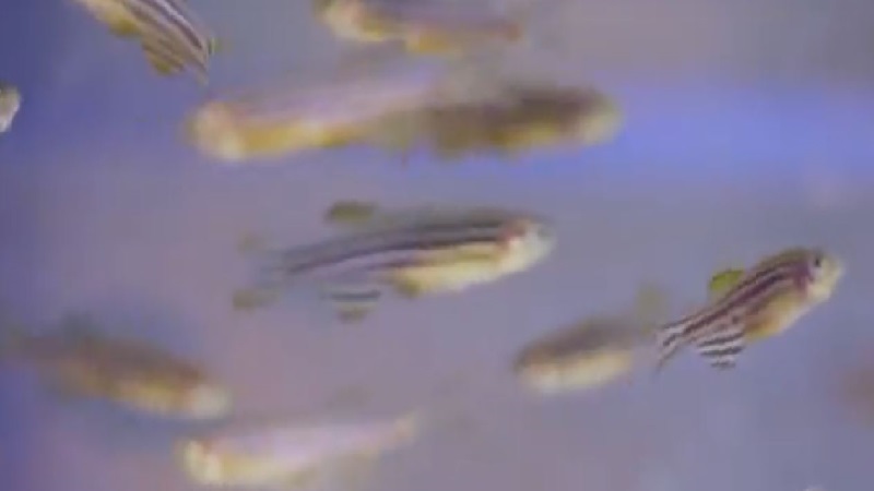
For the study, Blackshaw’s team focused on supportive cells in the back of the eye. In zebrafish, a standard laboratory model whose genome has been well defined, these cells, known as Müller glia, respond and repair the light-sensitive retina by growing new cells in the central nervous system called neurons. In addition to regrowing eye tissue, zebrafish’s regenerative abilities extend to other body parts, including fins, tails and some internal organs.
The retina is a good testing ground for mapping genetic activity, explains Blackshaw, because it contains structures common to other cells in the nervous system. In previous studies, moreover, scientists have found that the genetic networks in the retina are well conserved across species, so comparisons among fish, birds, mice and even humans are possible.
For the new experiments, the Johns Hopkins researchers created retinal injuries in zebrafish, chickens and mice. Then they used high-powered microscopes and a previously developed gene mapping tool to observe how the supportive Müller glia cells responded.
Blackshaw said the team was surprised to find, immediately after the injury, that the cells in each of the three species behaved the same way: They entered an “active state” characterized by the activation of specific genes, some of which control inflammation.
This active state, says Blackshaw, primarily helps to contain the injury and send signals to immune system cells to combat foreign invaders such as bacteria, or to clean up broken tissue.
Beyond that step, however, the species’ responses diverged.
In zebrafish, active Müller glia began turning on a network of transcription factors that control which genes are ‘on’ and ‘off.’ In the current experiment, the NFI transcription factors activated genes that are linked to cell maturity, sending the Müller glia cells back in developmental time to a more primitive state, which then allows them to develop into many different cell types. The Müller glia then “differentiated” into new cells to replace the ones lost to injury.
In contrast, the research team saw that chickens with damaged retinas activate only some of the transcription factor ‘gene control switches’ that are turned on in zebrafish. Thus, chickens have much less capability to create new Müller glia and other neurons in the eye following injury.
Finally, the researchers looked at the injury response in mice. Mice share the vast majority of their DNA with humans, and their eyes are similar to human eyes. The researchers found that injured Müller glia in mice remained in the first “active” state for several days, much longer than the eight to 12 hours that zebrafish are in this state, and yet never acquired the ability to make new neurons.
Müller glia in all three species also express high levels of nuclear factor I (NFI) transcription factors, but rapidly turn them off following injury. In mice, however, the NFI genes are turned back on soon thereafter, and actively block the Müller glia from generating neurons.
The researchers found, to their surprise, they say, that the same genes that allowed the zebrafish cells to regenerate were “primed and ready to go” in the mouse eye, but that the “on” transcription factor was never activated. Instead, the NFI factors actively block the cells’ regenerative potential.
Blackshaw suspects that animals with a higher potential to develop disease in brain and other neurological tissue may have lost this capability over evolutionary time to help protect and stabilize other brain cells. “For example, we know that certain viruses, bacteria and even parasites can infect the brain. It could be disastrous if infected brain cells were allowed to grow and spread the infection through the nervous system,” says Blackshaw.
Now equipped with a more detailed map of the cellular response to neuronal injury and regrowth, scientists may be able to find a way to activate the regenerative capabilities hidden in human DNA, Blackshaw says.
This work was a collaboration between investigators at Johns Hopkins, Notre Dame, Ohio State and the University of Florida supported by the Audacious Goals Initiative of the National Eye Institute. Other authors include Thanh Hoang, Fang Wang, Clayton Santiago, Lizhi Jiang, Cristian Saez, Fatemeh Rajaii, Trisha Parayil, Vickie Trinh, Dong Wong Kim, Guohua Wang and Jiang Qian of the Johns Hopkins University School of Medicine; Jie Wang of the Johns Hopkins University School of Medicine and West China Biomedical Big Data Center, West China Hospital, Sichuan University; Patrick Boyd, Manuela Lahne, Meng Jia, Leah Campbell and David Hyde of the University of Notre Dame; Sooyeon Yoo of the Johns Hopkins University School of Medicine and the Seoul National University Hospital; Casey Keuthan and John Ash of the University of Florida School of Medicine; Isabella Palazzo, Natalie Squires, Warren Campbell and Andy Fischer of the Ohio State University Wexner Medical Center.
This research was supported by the National Eye Institute (U01EY027267, R01EY024519, R01EY029548, K08EY027093, R01EY020560), the Hiller Family Endowment for Stem Cell Research at the University of Notre Dame and the Center for Zebrafish Research at the University of Notre Dame.
The authors declare no competing financial interest.