Because ear infections can be complicated to diagnose, otolaryngologist–head and neck surgeon James Clark occasionally sees patients who have been misdiagnosed with acute otitis media (AOM) or otitis media with effusion (OME). “After two or three courses of antibiotics, they come to a specialist like me because they are not improving,” says Clark.
To help alleviate the complexity of accurately identifying these conditions, in early 2019, Clark set out with Therese Canares, director of pediatric emergency medicine digital health innovation at Johns Hopkins, to develop a “smart” otoscope to diagnose AOM and OME using artificial intelligence. “I thought using technology might offer a solution,” says Clark.
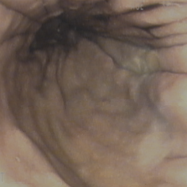
The clinicians joined a program at the Johns Hopkins Technology Innovation Center that matches researchers and health care providers with business, design and technical experts to develop software for improved patient care. Clark and Canares labeled 100 images of healthy and infected ear drums so that a computer using machine-learning could create an artificial intelligence algorithm. Once complete, initial testing showed the algorithm detects ear infections at the same sensitivity and specificity as a fellowship-trained otolaryngologist.
By October 2020, the team produced a prototype featuring a high-definition camera and screen, and wireless internet connectivity. After recording images of an eardrum, the smart otoscope sends the images to the cloud, where they are saved and then analyzed by the algorithm. A diagnosis is then sent back to the otoscope in real time and displayed on the screen. Clark says the technology may be beneficial for in-person appointments, telemedicine and urgent care settings.
“It can be difficult to explain to parents what’s going on in the ear without a picture,” he says. “The images can be shared with parents during the exam so they better understand why the child may or may not need an antibiotic.”
Alternatively, the technology could be used by a caregiver in the home to share high-resolution images with a physician in real time during a telehealth visit. In an urgent care setting, the device has potential to enhance speed and accuracy of diagnosis and treatment. “The clinician could prescribe an antibiotic right away if needed, and the patient could go home,” says Clark.
Since 2019, the team has secured $450,000 in funding. Most recently, they submitted a provisional patent application for the technology through Johns Hopkins Technology Ventures. The next step is gathering efficacy data on the prototype.
“The best thing about being at Hopkins is if you're interested in something, there's always someone you can call or email, and they will help you out,” says Clark. “There’s an openness to trying new technology across our health system, and that has been awesome.”