Advancing Care for Patients with Immunotherapy-Related Lung Injury
Imagine you are a patient with metastatic melanoma. After months of uncertainty, you finally begin immunotherapy, only to develop a relentless cough and shortness of breath a few weeks later. Tests reveal pneumonitis, a severe inflammation of the lungs triggered by the very treatment meant to save your life. The condition can cause lasting lung injury and dramatically alter the course of recovery.
Over the past decade, immunotherapy has revolutionized the treatment of advanced cancers, particularly melanoma, lung cancer, kidney cancer, bladder cancer and lymphoma, offering patients improved survival and, in some cases, long-term remission. Yet, these same therapies can also cause serious immune-related side effects. One of the most concerning is checkpoint inhibitor pneumonitis, an inflammatory complication of the lung that can occur in up to 10%–15% of patients receiving immunotherapy.
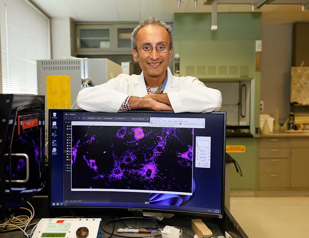
“Diagnosing and managing pneumonitis can be particularly challenging, and patients with moderate or severe symptoms typically require ICU levels of care,” says pulmonologist and critical care physician Karthik Suresh.
Johns Hopkins was one of the first institutions to identify and study this complication. The Department of Medicine collaborated with the Department of Oncology to establish the Immune-related Toxicity (Ir-tox) team, co-directed by rheumatologist Laura Cappelli and medical oncologist Aliyah Pabani. The team’s research showed that patients who developed pneumonitis had double the chance of dying, a finding that has been reproduced by other institutions.
Suresh has devoted his career to understanding and treating this condition. He and colleagues from the Ir-Tox team found that pneumonitis was more common and more deadly than initially reported. Working with colleagues in oncology and pulmonary medicine, Suresh was involved in the creation of one of the first Ir-Tox teams in the nation at Johns Hopkins, ensuring that patients with suspected pneumonitis could be rapidly evaluated and treated by a multidisciplinary group.
Uncovering the Mechanisms of Lung Injury
While pneumonitis was once a diagnosis of exclusion, research led by Suresh and his collaborators is beginning to define its biological underpinnings. In a 2019 study, his group demonstrated that patients with pneumonitis had abnormal populations of immune cells in their lungs, including pro-inflammatory macrophages.
More recently, together with colleagues from Massachusetts General Hospital, the University of Texas MD Anderson Cancer Center, the University of Miami, UZ Leuven (University Hospitals Leuven), Belgium, and Institute Gustave Roussy Cancer Institute in Paris, France, Suresh and colleagues have shown that macrophages in the lung produce high levels of a protein called CCL18. Elevated levels of CCL18 not only distinguish patients with pneumonitis from controls with accuracy, but also correlate with disease severity.
To test whether CCL18 was more than just a biomarker, Suresh and colleagues developed a novel mouse model. Mice engineered to overexpress CCL18 developed lung inflammation that closely mirrored human pneumonitis, strongly suggesting a causal role for this protein in promoting lung inflammation in immunotherapy-related pneumonitis.
These discoveries lay the groundwork for targeted therapies that could allow patients to continue lifesaving immunotherapy without being forced to discontinue treatment due to lung toxicity.
The Road Ahead
Suresh emphasizes that the pace of progress depends on sustained support. Federal grants from the National Institutes of Health and the American Thoracic Society Foundation have fueled this research, but philanthropic investment remains essential for recruiting patients, training junior investigators and accelerating the development of therapies. With additional resources, what has taken nearly a decade of painstaking work could be achieved in just a few years.
As immunotherapy continues to expand across cancer types, the need to predict, prevent and treat pneumonitis will only grow. The work led by Suresh and his collaborators represents a bridge from bench to bedside, turning basic discoveries into strategies that directly improve patient care.
This progress is only possible because of collaboration across Johns Hopkins, across institutions, and with patients and families who volunteer their time and tissue and blood samples for research. But moving from discovery to treatment requires more than science. It requires investment. Federal grants provide critical support, but philanthropy fills the gaps by funding personnel, expanding patient recruitment and sustaining the momentum of discovery.
By supporting this work, donors play a direct role in giving patients not only more time, but a better quality of life.
“Our goal is not just increasing survival, but also ensuring that our patients’ breathing and quality of life improves, and that they are able to continue getting therapies for treatment of their cancer,” Suresh says.
Together, we can turn cutting-edge research into safer treatments for patients everywhere.
For Clinicians Clinical Connection
Clinicians, discover the latest in research and clinical innovation from Johns Hopkins experts. Access educational videos, articles, CME courses and other resources from our world-renowned institution.