Exams We Offer: Mammogram
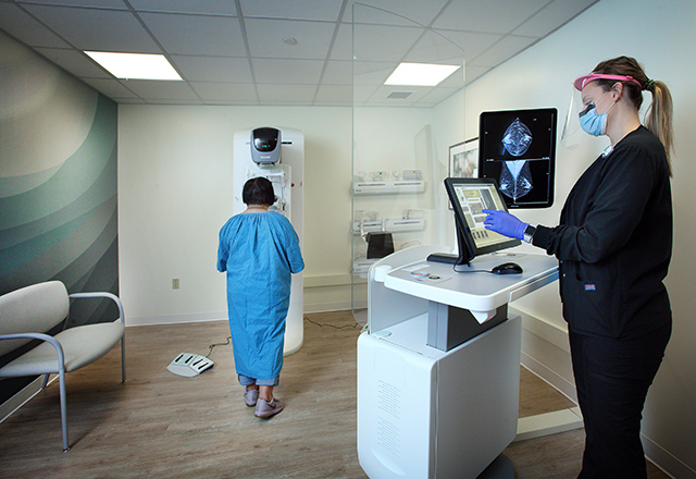
A mammogram is an X-ray of the breast used to screen for and diagnose breast cancer. At Johns Hopkins Medical Imaging sites, we exclusively offer 3-D mammography. 3-D mammography, or tomosynthesis, produces more detailed images of the breast tissue. Mammograms can also be used for a breast tissue or fluid biopsy.
Screening Mammograms
Annual screening mammograms are recommended for women who are 40 years or older, or for younger women with specific risk factors for breast cancer. You don’t have to have any signs or symptoms of a breast abnormality in order to receive a screening; they are used for the early detection of breast cancer and other breast health issues. Eighty percent of tumors found during a mammogram are benign. Screenings are also recommended for a period of time as follow-up care after breast cancer treatment.
Diagnostic Mammograms
You will be referred for this type of mammogram if you have an abnormality on your screening mammogram or if you have a breast mass or other breast change (found during a breast self-exam or by your physician). Diagnostic imaging may include mammograms with extra compression or magnification, mammograms shot from different angles or breast ultrasound.
Learn more about a mammogram exam in the Johns Hopkins Health Library.
Request An Appointment Request An Appointment

Schedule by phone

How do I prepare for a mammogram?
-
SCHEDULING: Breasts can be tender the week before and during menstruation, so try to schedule your mammogram for one to two weeks after your period starts. If you have breast implants, please notify the office when you schedule the exam.
Getting a mammogram too soon after your second dose of the coronavirus vaccine could result in a false positive and a callback due to temporarily swollen lymph nodes.
The Johns Hopkins Division of Breast Imaging supports the recommendation from the Society of Breast Imaging: When possible, and if it does not delay care your doctor recommends, you should schedule screening mammograms before your first dose of a COVID-19 vaccine or four to six weeks after the second dose.
PRECAUTIONS: If you are pregnant or think you may be pregnant, please check with your doctor before scheduling the exam. We will discuss other options with you and your doctor.
BREASTFEEDING: Please notify the technologist if you are currently breastfeeding.
PERSONAL HYGIENE: Do not use any deodorant, powder, lotion or perfume on the day of your exam.
CLOTHING: You must remove your clothing from the waist up and change into a patient gown and lock up all personal belongings. Please remove all piercings and leave all jewelry and valuables at home.
-
- You will be asked to remove your clothing from the waist up and any other clothing or jewelry that may interfere with the exam.
- You will stand in front of the mammography machine and one breast will be placed on the X-ray plate.
- A separate flat plate will be brought down on top of the breast to compress it against the X-ray plate. You may feel some discomfort or pressure on your breasts during this X-ray process. This part of the process should only last for a few minutes.
Learn more about what happens during a mammogram exam in the Johns Hopkins Health Library.
-
There is typically no special type of care following a mammogram. However, your health care provider may give you additional instructions depending on your specific health condition.
What to Expect During Your First Mammogram
A mammogram is an important step in taking care of yourself and your breasts but not knowing what to anticipate can be stressful. Join Monica for her first mammogram experience with Johns Hopkins Medical Imaging to learn more about how to prepare for a mammogram and what to expect during and after the exam.
Learn more about mammograms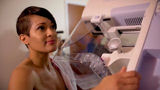
Why Choose Johns Hopkins Medical Imaging?
Physician Experts

Top Radiology Department
State-of-the-Art Technology

Your Safety Is Always Our Priority

Specialty Technologists

Patient Care
