Research Lab Results
-
Laboratory for Computational Motor Control
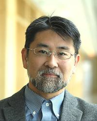
The Laboratory for computational Motor Control studies movement control in humans, including healthy people and people with neurological diseases. We use robotics, brain stimulation and neuroimaging to study brain function. Our long-term goals are to use mathematics to understand: 1) the basic function of the motor structures of the brain including the cerebellum, the basal ganglia and the motor cortex; and 2) the relationship between how our brain controls our movements and how it controls our decisions. -
Leucker Laboratory
The Leucker Laboratory is a translational cardiovascular research program dedicated to understanding the mechanisms underlying residual atherosclerotic cardiovascular disease risk and developing innovative strategies for cardiovascular prevention. Our work integrates human vascular biology, advanced cardiovascular imaging, epidemiology, and clinical trials to uncover the pathways linking inflammation, lipoproteins, endothelial dysfunction, and cardiometabolic disease to cardiovascular outcomes.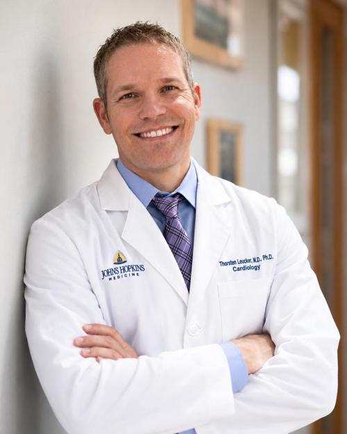
Principal Investigator
-
Nicholas Dalesio Lab
Research in the Nicholas Dalesio Lab is currently examining pre-surgical predictors of post-surgical respiratory complications in children with obstructive sleep apnea and sleep-disordered breathing; the impact of anesthesia and pharmacological agents on upper airway physiology; and techniques for pediatric airway imaging.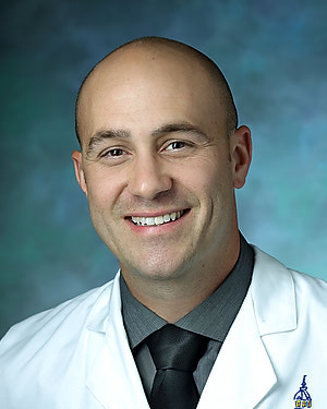
-
Nicholas Flavahan Lab
The Nicholas Flavahan Lab primarily researches the cellular interactions and subcellular signaling pathways that control normal vascular function and regulate the initiation of vascular disease. We use biochemical and molecular analyses of cellular mediators and cell signaling mechanisms in cultured vascular cells, while also conducting physiological assessments and fluorescent microscopic imaging of signaling systems in isolated blood vessels. A major component of our research involves aterioles, tiny blood vessles that are responsible for controlling the peripheral resistance of the cardiovascular system, which help determine organ blood flow. -
O'Connor Lab
How do brain dynamics give rise to our sensory experience of the world? The O'Connor lab works to answer this question by taking advantage of the fact that key architectural features of the mammalian brain are similar across species. This allows us to leverage the power of mouse genetics to monitor and manipulate genetically and functionally defined brain circuits during perception. We train mice to perform simple perceptual tasks. By using quantitative behavior, optogenetic and chemical-genetic gain- and loss-of-function perturbations, in vivo two-photon imaging, and electrophysiology, we assemble a description of the relationship between neural circuit function and perception. We work in the mouse tactile system to capitalize on an accessible mammalian circuit with a precise mapping between the sensory periphery and multiple brain areas. Our mission is to reveal the neural circuit foundations of sensory perception; to provide a framework to understand how circuit dysfunction causes mental and behavioral aspects of neuropsychiatric illness; and to help others fulfill creative potential and contribute to human knowledge. -
Inoue Lab
Complexity in signaling networks is often derived from co-opting one set of molecules for multiple operations. Understanding how cells achieve such sophisticated processing using a finite set of molecules within a confined space--what we call the ""signaling paradox""--is critical to biology and engineering as well as the emerging field of synthetic biology. In the Inoue Lab, we have recently developed a series of chemical-molecular tools that allow for inducible, quick-onset and specific perturbation of various signaling molecules. Using this novel technique in conjunction with fluorescence imaging, microfabricated devices, quantitative analysis and computational modeling, we are dissecting intricate signaling networks. In particular, we investigate positive-feedback mechanisms underlying the initiation of neutrophil chemotaxis (known as symmetry breaking), as well as spatio-temporally compartmentalized signaling of Ras and membrane lipids such as phosphoinositides. In parallel, we also try to understand how cell morphology affects biochemical pathways inside cells. Ultimately, we will generate completely orthogonal machinery in cells to achieve existing, as well as novel, cellular functions. Our synthetic, multidisciplinary approach will elucidate the signaling paradox created by nature. -
Michael Caterina Lab
The Caterina lab is focused on dissecting mechanisms underlying acute and chronic pain sensation. We use a wide range of approaches, including mouse genetics, imaging, electrophysiology, behavior, cell culture, biochemistry and neuroanatomy to tease apart the molecular and cellular contributors to pathological pain sensation. A few of the current projects in the lab focus on defining the roles of specific subpopulations of neuronal and non-neuronal cells to pain sensation, defining the role of RNA binding proteins in the development and maintenance of neuropathic pain, and understanding how rare skin diseases known as palmoplantar keratodermas lead to severe pain in the hands and feet.
-
Mukherjee Lab
The Mukherjee Cardiovascular Innovations Lab harnesses cutting-edge imaging techniques to explore cardiovascular manifestations and enhance the screening, early detection, and prediction of adverse clinical events across a broad range of autoimmune diseases. -
Molecular Oncology Laboratory
Our Molecular Oncology lab seeks to understand the genomic wiring of response and resistance to immunotherapy through integrative genomic, transcriptomic, single-cell and liquid biopsy analyses of tumor and immune evolution. Through comprehensive exome-wide sequence and genome-wide structural genomic analyses we have discovered that tumor cells evade immune surveillance by elimination of immunogenic mutations and associated neoantigens through chromosomal deletions. Additionally, we have developed non-invasive molecular platforms that incorporate ultra-sensitive measurements of circulating cell-free tumor DNA (ctDNA) to assess clonal dynamics during immunotherapy. These approaches have revealed distinct dynamic ctDNA and T cell repertoire patterns of clinical response and resistance that are superior to radiographic response assessments. Our work has provided the foundation for a molecular response-adaptive clinical trial, where therapeutic decisions are made not based on imaging but based on molecular responses derived from liquid biopsies. Overall, our group focuses on studying the temporal and spatial order of the metastatic and immune cascade under the selective pressure of immune checkpoint blockade with the ultimate goal to translate this knowledge into “next-generation” clinical trials and change the way oncologists select patients for immunotherapy.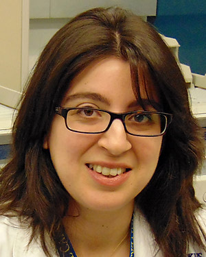
-
The Mumm Lab
The research conducted in the Mumm Lab (Dept. of Ophthalmology, Wilmer Eye Institute) is focused on understanding how neural circuits are formed, how they function, and how they can be regenerated, to develop new therapies for retinal regeneration. Toward that end, we investigate the development, function, and regeneration of disease-relevant neurons and neural circuits responsible for vision. An emphasis is placed on translating what can be learned in regenerative model systems to develop novel therapies for stimulating dormant regenerative capacities in humans, Therefore, we apply what we learn from a naturally regenerative species, the zebrafish, toward the development of novel therapies for restoring visual function to patients. We place an emphasis on unique perspectives zebrafish afford to biological studies, such as in vivo time-lapse imaging of cellular behaviors and cell-cell interactions, and high-throughput chemical and genetic screening. We have pioneered several technologies to support this work including multicolor imaging of neural circuit formation, a selective cell ablation methodology, and a quantitative high-throughput phenotypic screening platform. Together, these approaches are providing novel insights into how the degeneration and regeneration of discrete retinal cell types is controlled.