Research Lab Results
-
Kathryn Carson Lab
The Kathryn Carson Lab investigates ways to improve medical research, particularly in the areas of brain and thyroid cancer, Alzheimer’s disease, atherosclerosis, hypertension, HIV and lupus. Our team seeks to help researchers optimize their studies through better study design, protocol and grant writing, data cleaning and analysis, and publication writing. We work with investigators from a wide range of departments through the Johns Hopkins Institute for Clinical and Translational Research. -
Kathleen Cullen Lab
We are continually in motion. This self-motion is sensed by the vestibular system, which contributes to an impressive range of brain functions, from the most automatic reflexes to spatial perception and motor coordination. The objective of Dr. Cullen's lab's research program is to understand the mechanisms by which self-motion (vestibular) information is encoded and then integrated with signals from other modalities to ensure accurate perception and control of gaze and posture. Our studies investigate the sensorimotor transformations required for the control of movement, by tracing the coding of vestibular stimuli from peripheral afferents, to behaviorally-contingent responses in central pathways, to the readout of accurate perception and behavior. Our experimental approach is multidisciplinary and includes a combination of behavioral, neurophysiological and computational approaches in alert behaving non-human primates and mice. Funding for the laboratory has been and is provided by the Canadian Institutes for Health Research (CIHR), The National Institutes of Health (NIH), the National Sciences and Engineering Research Council of Canada (NSERC), FQRNT / FQRSC (Quebec). -
King-Wai Yau Laboratory
The King-Wai Yau Laboratory is interested in the area of sensory transduction. Specifically, we study visual transduction, the process by which the sense of vision is initiated. In our eyes, we have two primary photoreceptors-rods and cones-to absorb light and convert light into electrical signal, which is then transmitted to the brain for image formation. Rods are extremely sensitive and responsible for night vision, but cones are about 100-fold less sensitivity and responsible for daylight vision. We are studying the cellular and molecular details underlying rod and cone phototransduction, aiming to understand the mechanism of rod-cone difference. Our ultimate goal is to convert rods into cones, or cones into rods to rescue human vision loss. -
Kristina Nielsen Laboratory
The Kristina Nielsen Laboratory investigates neural circuits in the visual cortex that are responsible for encoding objects to understand how the visual system performs object recognition. We aim to reveal the fine-scale organization of neural circuits, with an emphasis on higher-level visual areas. We use two-photon microscopy to perform high-resolution functional imaging of visual areas in the non-human primate. We also investigate how the function of higher visual areas changes over the course of brain development in ferrets, by measuring the activity of single neurons in these areas, as well as determining the animal's visual capabilities at various developmental stages. In both types of investigations, we also rely on detailed anatomical techniques to precisely observe how the function of neuronal circuits is related to their structure. -
Kenneth W. Kinzler Laboratory
Dr. Kinzler’s laboratory has focused on the genetics of human cancer. They have identified a variety of genetic mutations that underlie cancer, including mutations of the APC pathway that appear to initiate the majority of colorectal cancers and IDH1/2 mutations that underlying many gliomas. In addition, they have developed a variety of powerful tools for analysis of expression and genetic alterations in cancer. Most recently, they have pioneered integrated whole genome analyses of human cancers through expression, copy number, and mutational analyses of all the coding genes in several human cancer types including colorectal, breast, pancreatic and brain. The identification of genetic differences between normal and tumor tissues provide new therapeutic targets, new opportunities for the early diagnosis of cancer, and important insights into the neoplastic process.
-
Lee Martin Laboratory
In the Lee Martin Laboratory, we are testing the hypothesis that selective vulnerability--the phenomenon in which only certain groups of neurons degenerate in adult onset neurological disorders like amyotrophic lateral sclerosis and Alzheimer's disease--is dictated by brain regional connectivity, mitochondrial function and oxidative stress. We believe it is mediated by excitotoxic cell death resulting from abnormalities in excitatory glutamatergic signal transduction pathways, including glutamate transporters and glutamate receptors as well as their downstream intracellular signaling molecules. We are also investigating the contribution of neuronal/glial apoptosis and necrosis as cell death pathways in animal (including transgenic mice) models of acute and progressive neurodegeneration. We use a variety of anatomical and molecular neurobiological approaches, including neuronal tract-tracing techniques, immunocytochemistry, immunoblotting, antipeptide antibody production, transmission electron microscopy and DNA analysis to determine the precise regional and cellular vulnerabilities and the synaptic and molecular mechanisms that result in selective neuronal degeneration.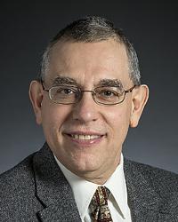
-
Neuroimaging and Modulation Laboratory (NIMLAB)
The neuroimaging and Modulation Laboratory (NIMLAB) investigates neural correlates of cognition and behavior using neuroimaging methods such as functional magnetic resonance imaging (fMRI) and neuromodulation techniques such as transcranial magnetic stimulation (TMS). We are looking in depth at the contributions of the cerebellum and cerebro-cerebellar circuits to cognition; the effects of chronic heavy alcohol consumption on cognition and brain activation underlying cognitive function; how aging in humans affects neural systems that are important for associative learning and stimulus awareness; and the integration of transcranial magnetic stimulation with functional MRI. -
Neuro-Vestibular and Ocular Motor Laboratory
In our laboratory we study the brain mechanisms of eye movements and spatial orientation. -How magnetic stimulation through transcranial devices affects cortical brain regions -Neural mechanisms underlying balance, spatial orientation and eye movement -Mathematical models that describe the function of ocular motor systems and perception of spatial orientation -Short- and long-term adaptive processes underlying compensation for disease and functional recovery in patients with ocular motor, vestibular and perceptual dysfunction Developing and testing novel diagnostic tools, treatments, and rehabilitative strategies for patients with ocular motor, vestibular and spatial dysfunction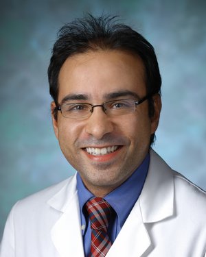
-
Neuro-Oncology Surgical Outcomes Laboratory
Directed by Debraj “Raj” Mukherjee, MD, MPH, the laboratory focuses on improving access to care, reducing disparities, maximizing surgical outcomes, and optimizing quality of life for patients with brain and skull base tumors.
The laboratory achieves these aims by creating and analyzing institutional and national databases, developing and validating novel patient-centered quality of life instruments, leveraging machine learning and artificial intelligence platforms to risk-stratify vulnerable patient populations, and designing novel surgical trials to push the boundaries of neurosurgical innovation.
Our research also investigates novel approaches to improve neurosurgical medical education including studying the utility of video-based surgical coaching and the design of new operative instrumentation.
-
Vestibular and Ocular motor Research Laboratory
Our research is directed toward how the brain controls the movements of the eyes (including eye movements induced by head motion) using studies in normal human beings, patients and experimental animals. The focus is on mechanisms underlying adaptive ocular motor control. More specifically, what are mechanisms by which the brain learns to cope with the changes associated with normal development and aging as well as the damage associated with disease and trauma? How does the brain keep its eye movement reflexes properly calibrated? Our research strategy is to make accurate, quantitative measures of eye movements in response to precisely controlled stimuli and then use the analytical techniques of the control systems engineer to interpret the findings. Research areas: 1) learning and compensation for vestibular disturbances that occur either within the labyrinth or more centrally within the brain, 2) the mechanisms by which the brain maintains correct alignment of the eyes to prevent diplopia and strabismus, and 3) the role of ocular proprioception in localizing objects in space for accurate eye-hand coordination.