Covid-19 Story Tip: Researchers May Have Unclouded the Mystery of COVID-19 ‘Brain Fog’
02/24/2021
People who survive a severe case of COVID-19 have reported a variety of lingering aftereffects, but perhaps none as unusual as what has been popularly called “brain fog.” Clinically known as dysexecutive syndrome, the condition is a COVID-19-kindled delirium, initially experienced by patients — mostly older — while sick as a state of confusion and impaired awareness. It then often stays on after recovery to torment as persistent cognitive sluggishness. Unfortunately, the origin of brain fog has remained unclear.
Now, pathologists at Johns Hopkins Medicine in Baltimore and Brigham and Women’s Hospital in Boston have found evidence that large bone marrow cells known as megakaryocytes may be responsible for the brain fog. They suggest that megakaryocytes migrate to the brain in a journey precipitated by the destructive activity of SARS-CoV-2, the virus that causes COVID-19.
There, the researchers believe, the out-of-place cells may reduce or completely block the flow of nourishing blood through individual capillaries in the cerebral cortex — the area of the brain where most information processing occurs. Such capillary occlusions, they say, could lead to neurological impairment.
The team’s findings are reported in a research letter published Feb. 12 in JAMA Neurology.
Megakaryocytes are the cells responsible for production of platelets —blood components that are necessary for clotting and wound repair.
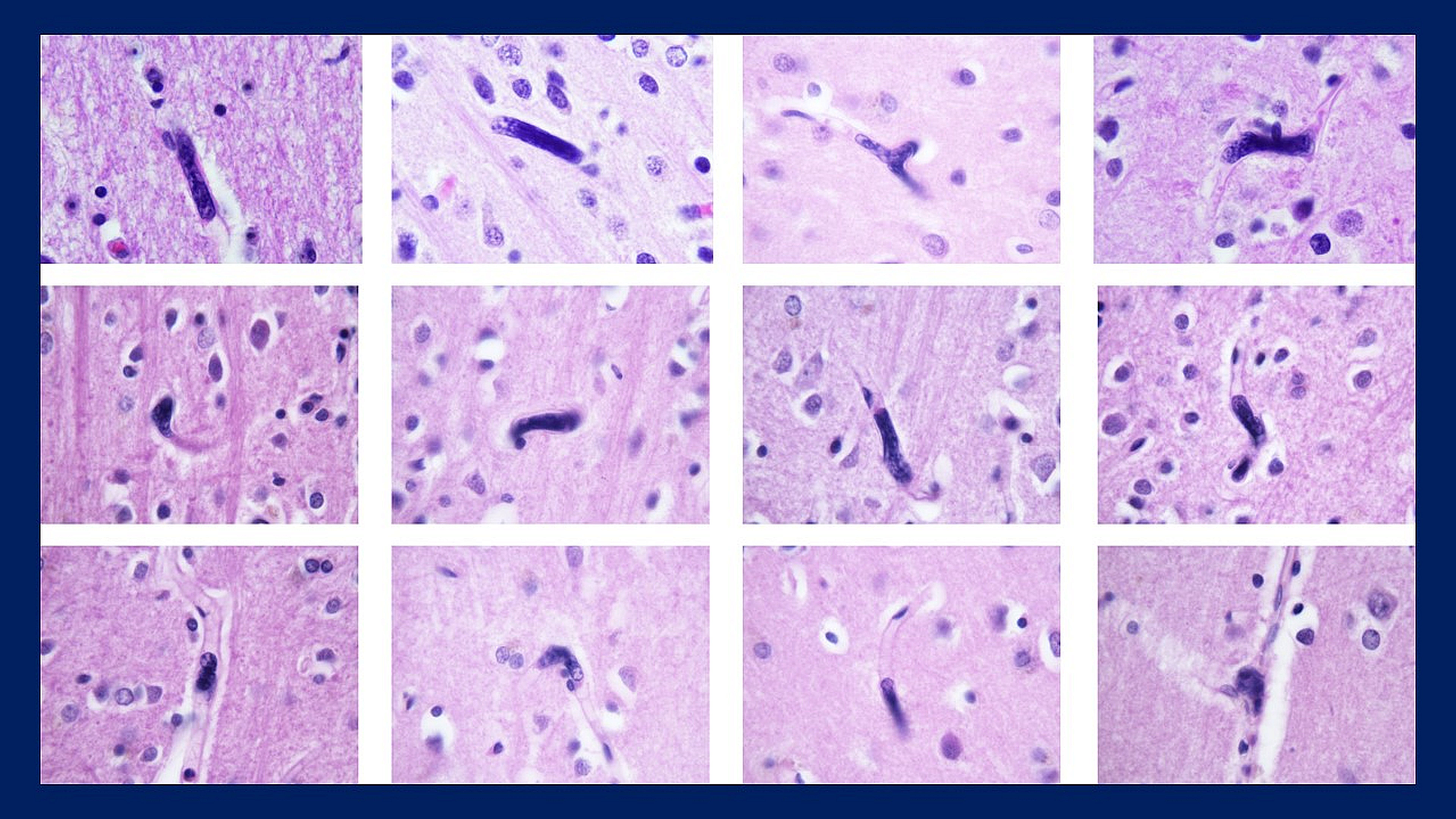
Interestingly, the researchers say, a scientific literature search they conducted indicates this may be the first time that megakaryocytes have been found in blood vessels in the human brain.
The researchers evaluated brain tissue from autopsies of 15 patients who died of COVID-19. In five of the samples, they discovered large cell nuclei resembling those of megakaryocytes in the cortical capillaries. Immunohistochemical testing confirmed that the nuclei were those of megakaryocyte immigrants from bone marrow.
No megakaryocyte nuclei were found in post-mortem brain tissues from decedents who were negative for the COVID-19 coronavirus and served as the study’s control group.
What isn’t clear is how the COVID-19-associated megakaryocytes are signaled to leave the bone marrow or how they can navigate the fine network of lung blood vessels on their way to the brain, says David Nauen, M.D., Ph.D., study lead author and assistant professor of pathology at the Johns Hopkins University School of Medicine.
“We suspect that SARS-CoV-2 damages lung tissue, leading to the release of chemical signals that induce the megakaryocytes to travel there from the bone marrow,” says Nauen. “When that happens, these large cells somehow find a way to pass through the lung capillaries and get to the brain.”
“We don’t yet know if the megakaryocytes we found in the brain are just the result of blood flow carrying them there or if a specific change occurs in the brain vessels that trap them,” he adds.
Nauen says the researchers plan next to characterize what happens in the brain tissue and cortical capillaries during severe cases of COVID-19 to better understand how megakaryocytes are signaled and recruited.
“Because standard brain autopsy sections represent only a minute portion of the cerebral cortex, the actual numbers of these cells in the brain as a result of COVID-19 could be considerable — as could their potentially negative neurological impacts on those who survive,” says Nauen.
Nauen is available for comment about these research findings.