Johns Hopkins Fundamentals: Top 9 Science Images Of 2018
12/20/2018

Jan. 1 marked the launch of a visual storytelling campaign for science stories from Johns Hopkins Medicine. Here are the top 9 images from this year from our Instagram.
Follow @JHM.Fundamentals on Instagram for new science images each week. For more information about the images or the research they represent, contact Rachel Butch: [email protected]; 410-955-8665.
Click the images to view the Instagram posts.
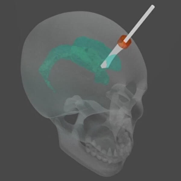 |
Open your mind (or brain) to this ultrasonic probe. It may look scary, but this miniaturized tool is helping surgeons remove tumors from the brains of their patients. The probe acts like a magnifying lens in the sun, but instead of burning paper, the ultrasound device focuses sound into powerful waves that precisely burn away cancer cells. When placed inside pools of cerebrospinal fluid within the skull (shown in green), this probe minimizes the risk of infection and blood loss to patients undergoing this delicate procedure. |
 |
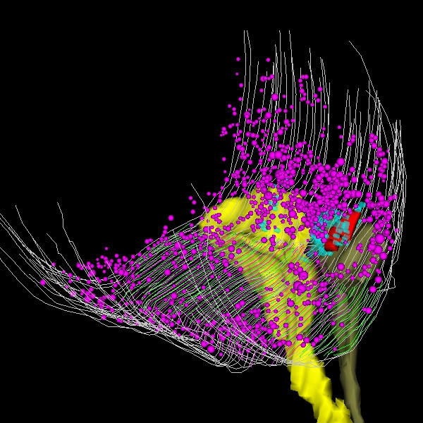
The human brain is loaded with some 100 billion nerve cells, each connecting to thousands of others, resulting in around 100 trillion connections. Mapping those connections could enable scientists to decipher the causes of neurological disease and mental illness. It's an immense, daunting task. The best way to tackle it? Huge amounts of data, say researchers. Biomedical engineer Josh Vogelstein, creates images like this one from data collected from brain scans to help researchers navigate the labyrinthine pathways of the brain. |
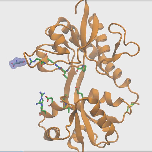 |
Highway or local roads? Johns Hopkins scientists have plotted the path that brain signals take to connect to neurons. They used supercomputers to create this model of how signaling chemicals fit into receptors on neurons. Biophysicist and biochemist Albert Lau hopes to shed light on the physics of the chemical’s pathway, as well as the speed of nerve cell communications using models like this one. Check out the news release: Johns Hopkins Scientists Chart How Brain Signals Connect to Neurons |
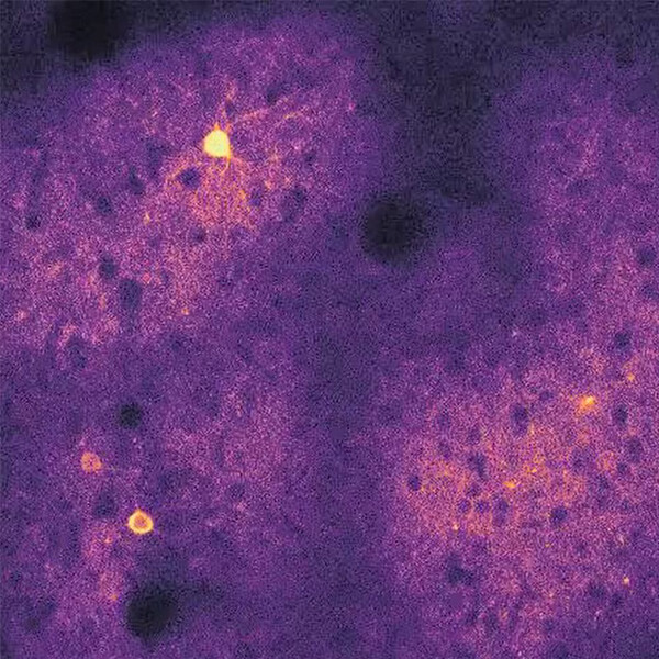 |
The “fireworks” shown here are courtesy of one of our neuroscientists, Rick Huganir. Each flash is a burst of energy traveling through a neuron. Neurons in your brain form a vast constellation and respond to different aspects of visual experience, allowing us to perceive the world around us. Huganir hopes to use images like this one to learn how individual excitatory neurons tune in to different visual input. |
 |
Copper is essential to human biology and makes many processes — from the formation of color in your hair and eyes to new blood vessels — possible. Imbalances in the mineral have been associated with several neurological disorders, depression and changes in sleep. But don’t start sucking on pennies just yet — copper deficiency is not a common problem. A diet incorporating vegetables, nuts and even chocolate usually contains enough copper to keep you happy and healthy. Check out the news release: Low Copper Levels Linked to Fatter Fat Cells |
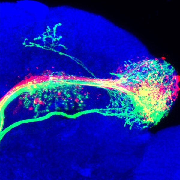 |
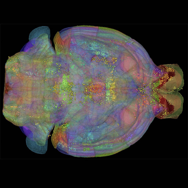
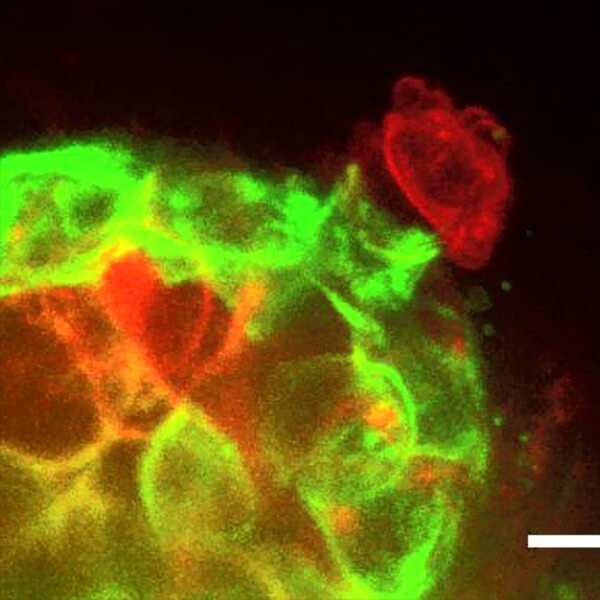
| Neuroscientist Chris Potter uses images like this fruit fly brain to find out more about how the insect’s sense of smell. Green, red and yellow light up neurons reacting to different scents in the air. |
 |
Can you hear when someone whispers? Thank your outer hair cells, the unsung heroes of the inner ear. The cells amplify sound waves to help you hear quiet noises, and they are inhibited by the brain to protect you from loud ones. Paul Fuchs’ lab created this model of the bottom of an outer hair cell using an imaging technique called electron micrography. |
 |
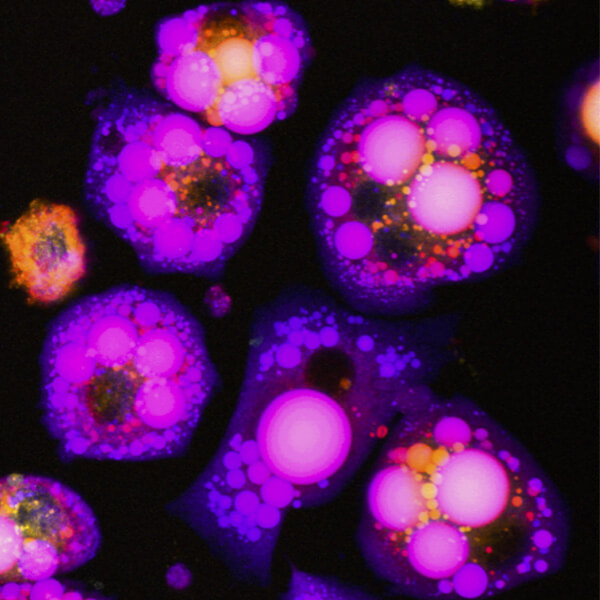
The colorful world of cancer drug discovery. Melanoma is the most deadly form of skin cancer, with nearly 9,000 deaths reported each year. That is why finding a treatment that can target these cells has become the focus of Nick Low’s postdoctoral work. Through his research, Low found ways to throw a wrench in melanoma’s cellular machinery. His lab discovered a small molecule that destroys the enzyme that allows these cancer cells to turn their DNA into proteins, called RNA polymerase. By investigating how these cellular processes are regulated, Low hopes to develop new cancer treatments. |
 |
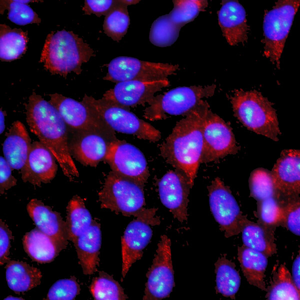
The cells surrounding your breast duct are more like a soccer team than a castle wall, researcher Andrew Ewald says. When there’s a tumor in the breast duct, these barrier cells don’t just stand in the way of cancer cells trying to escape. They can actively “play defense”, moving to block and grab the cells. Check out the news release: Scientists Discover a Dynamic Cellular Defense Against Breast Cancer Invasion |