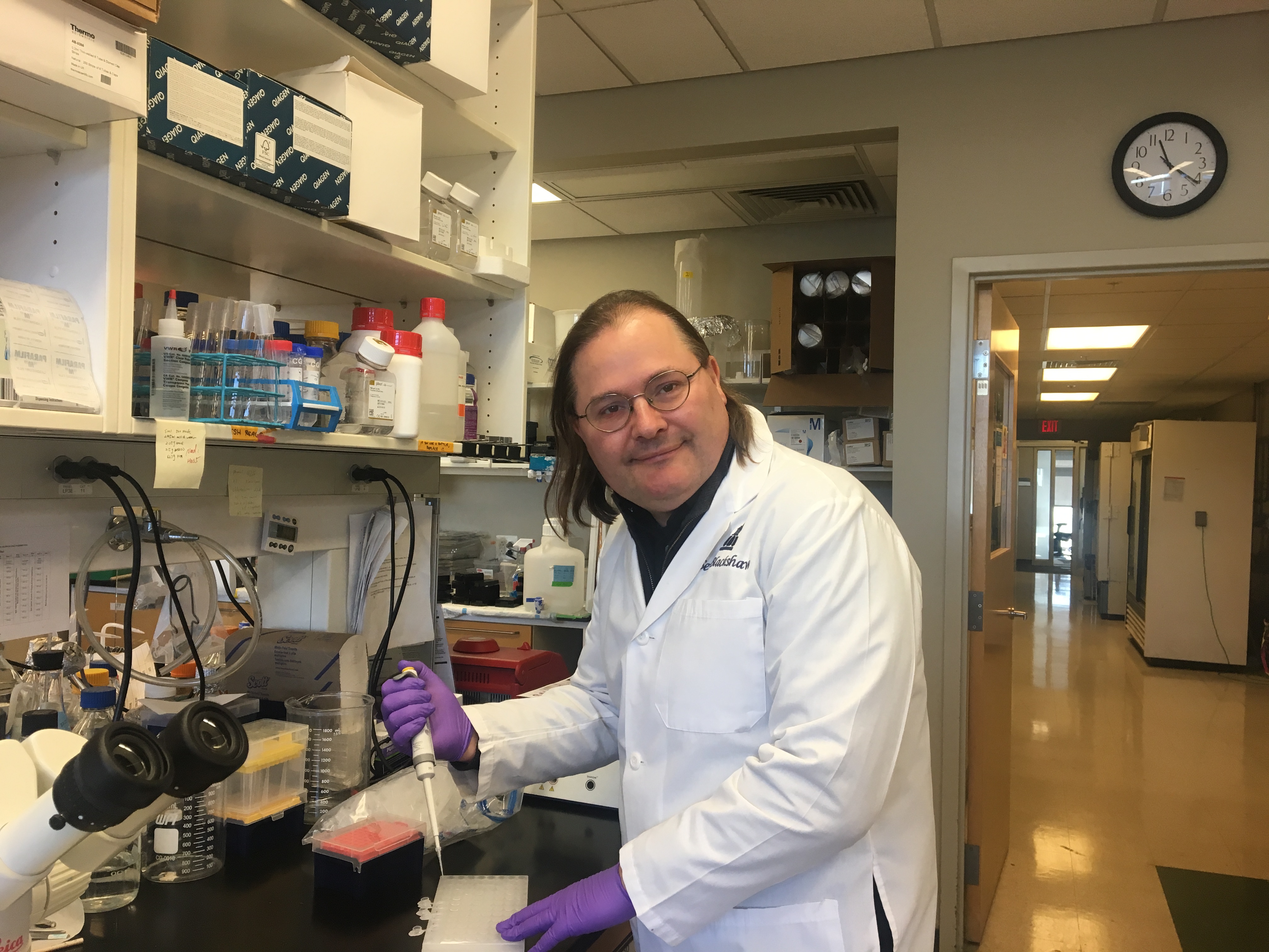
In mammals, retinal neurons are lost forever following injury, meaning that damage to these cells from trauma and diseases such as macular degeneration and glaucoma is permanent. That isn’t the case, however, for all vertebrates. Zebrafish, for example, are able to regenerate retinal neurons from a type of non-neuronal support cell known as Müller glia following injury. Now a team led by Seth Blackshaw, a professor of neuroscience, neurology and ophthalmology at the Johns Hopkins University School of Medicine, has discovered that mammals retain this ability, but that during the course of evolution, it was — in the genetic sense — switched off.
Four years ago, Blackshaw and colleagues from The Johns Hopkins University, the University of Notre Dame, Ohio State University and the University of Florida embarked on a study to determine what happens in retinal glia in multiple different models of injury. They compared what happens in genes that cannot regenerate (mice), that can regenerate well (zebrafish) and that have very limited ability to regenerate (newly hatched chicks).
Studying models of both inner and outer retinal injury, from the time before injury occurred to the time glia had fully reprogrammed, they observed which genes were activated or repressed at a specific time point and how those genes are themselves regulated by transcription factors, which are proteins responsible for turning genes on and off.
By profiling everything that happened in the glia over the course of injury, the team was able to reconstruct those dynamic regulatory networks and see the common ways glia respond to injury, what is specific to individual species and how that correlates with the ability to regenerate.
Following injury, glia from all species enter an activated state. They express common sets of genes. But what happens after that differs considerably, says Blackshaw. In fish, there’s a set of gene regulatory networks that is switched on when cells enter this activated state, so the glia quickly reprogram to become neurogenic progenitors — cells with the capacity to regenerate neurons. In chickens, a lot of the glia revert to the resting state, but some then go on to reprogram. But in mice, none of them reprogram. They get to the activated state and stop. “Interestingly, they actually start to transiently express some genes associated with reprogramming, but those quickly get shut off,” Blackshaw says.
In the mice, Blackshaw and his colleagues discovered a series of inhibitory gene regulatory networks overlying the gene regulatory network that promotes reprogramming, which prevents the mice glia from reprogramming in response to injury. When they disrupted genes in the inhibitory gene regulatory networks, the mouse retinal glia acquired the ability to proliferate and generate neurons.
An Evolutionary Trade-Off
The researchers posit that the networks changed over the course of evolution. So why did mammals lose the ability to reprogram injured glia? That’s the big question, says Blackshaw, who hypothesizes that they evolved as a means of resisting infection.
Instead of reprogramming following injury, mammalian glia enter a prolonged activated state called gliosis, in which they swell up and stiffen, forming a protective physical barrier around the focal site of injury. They then kill severely injured cells before releasing anti-inflammatory molecules to protect less severely damaged neurons. Blackshaw says this isn’t specific to mammals. Adult birds have also lost the ability to regenerate, as have certain cold-blooded animals. And one thing that warm-blooded species like mammals and birds tend to have in common, he says, is a higher parasite load.
“Traditionally, parasite load might be considered a relatively minor problem as compared to, say, the problem of macular degeneration, but I think COVID-19 has demonstrated why the immune system wants to do what it can to prevent pathogens from getting into the central nervous system,” says Blackshaw. He believes his hypothesis is testable by genetically disrupting the ability of zebrafish Müller glia to reprogram and then seeing if they become more susceptible to infection.

The team has had some success in reprogramming mouse Müller glia by knocking out some of the genes that normally prevent reprogramming from occurring after injury. They’ve been able to generate certain inner retinal neurons, but not those typically affected in retinal disease. Now they’re trying to make the process more efficient by targeting other genes in the network. They’re also trying to get the reprogrammed Müller glia to make new photoreceptors and ganglion cells — the neurons lost in macular degeneration and glaucoma, respectively.
Because the ability to regenerate glia occurs in the brain in the same way it does in the retina, this work has broader implications for understanding other neurodegenerative disorders and neural injuries. In addition, Blackshaw points out that the loss of the ability to make cells regenerate new neurons goes hand in hand with the loss of the ability to regenerate axons, which has implications for things like spinal cord injury. “Many of the same genes are expressed in glial cells elsewhere in the brain, so I suspect that if these basic mechanisms are found in glial cells elsewhere in the central nervous system, we may be looking at useful strategies for treating a whole host of neurodegenerative disorders and neural injuries,” says Blackshaw.
Thirteen additional Johns Hopkins affiliates from the Department of Neurology, the Department of Ophthalmology, the Institute for Cell Engineering, the Kavli Neuroscience Discovery Institute and the Solomon H. Snyder Department of Neuroscience contributed to this study.
Read more about the group’s work, which was published in the journal Science.