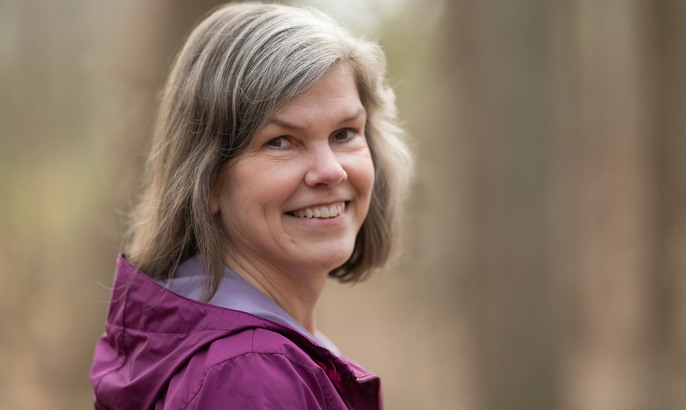
In February of 2020, Tori Banks, a 52-year-old copy editor from Perry Hall, Maryland, started having periodic headaches. At first, she remembers, they were mild — but over the next few weeks, they morphed into something different entirely. On Super Bowl Sunday that year, she was gripped by a headache so incapacitating that a friend who’s a nurse told her afterward that she should go straight to the emergency room if another one struck. Days later, Banks did just that, where staff at Johns Hopkins Bayview Medical Center’s emergency department ran a gamut of tests, including a CT scan and MRI of her head.
“They came in and said right away, ‘There’s something in there,’” says the mother of two. “The CT scan showed that I didn’t have any bleeding in my brain, which was good, but the MRI revealed that I had a brain tumor.”
Within days, Banks had an appointment with Johns Hopkins neurosurgeon Debraj “Raj” Mukherjee, who worked with his colleagues to plan surgery to remove Banks’ tumor, a mass on the right side of her head. However, he also communicated carefully with Banks to accommodate her personal goals into the surgical plan. Because of the tumor’s precarious position near her optic pathway, surgery to remove it had a high chance of partially blinding her — a consequence that could be extremely detrimental for Banks’ career and impede her ability to enjoy family events like her daughter’s college graduation. So the surgeons left a tiny segment of tumor in place (a small piece that they believed would be eradicated with chemotherapy and radiation after surgery), preserving Banks’ eyesight and allowing her to continue doing the things most important to her.
Banks’ experience represents a notable shift in the field of brain cancer research and treatment today, a field that too often has focused on extending life at the expense of preserving the quality of that life, says Mukherjee. At Johns Hopkins, an increasing number of studies — including many led by Mukherjee and colleagues throughout Johns Hopkins’ Departments of Neurosurgery and Neurology, and the Division of Neuroradiology — are now focused on ensuring that patients with brain cancer can live as comfortably as possible and remain able to participate in and enjoy the activities they care about most.
Together, these three neuro groups tag-team to care for around 2,500 adult and pediatric patients with brain cancer each year, providing the oncology staples of surgery, chemotherapy and radiation, along with addressing the array of other needs that patients with brain cancer encounter along their journey. The data and advances derived from their research are helping those treated at Johns Hopkins and beyond preserve the function and meaning that are key to living the best lives they can.
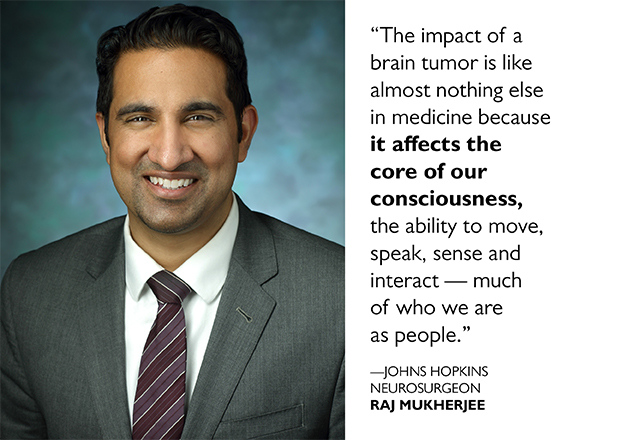
“The impact of a brain tumor is like almost nothing else in medicine because it affects the core of our consciousness, the ability to move, speak, sense and interact — much of who we are as people,” Mukherjee says. “Living longer after diagnosis is very important, but living as well as possible is equally significant.”
Benefits of Liquid Biopsies
The brain’s role as neurological master controller makes it especially challenging for doctors to treat brain tumors without negatively affecting quality of life, explains Mukherjee’s colleague, neurosurgeon Chetan Bettegowda.
Depending on a tumor’s location and size, surgery or radiation could affect a variety of factors considered essential to living well, such as mobility, any of the senses, memory and cognitive function.
Also factoring into the treatment challenge is the type of brain tumor being treated, which can range from a benign tumor to aggressive and deadly tumor types including Banks’ ultimate diagnosis of glioblastoma. The fast-acting glioblastoma is the most common primary brain malignancy in adults. With the average survival time for glioblastoma only 12 to 18 months after diagnosis, making the most of that time for patients and their families is especially important.
Regardless of tumor type, says Bettegowda, catching tumors early and monitoring the results of therapy are pivotal to preserving quality of life: The earlier patients can be diagnosed and receive effective treatment, the more function can be preserved. Toward this goal, the vast majority of research in his lab focuses on liquid biopsies, tests that he and his colleagues are developing to sense the presence of tumor-specific DNA, proteins and other biomarkers. Bettegowda is widely considered a leader in this field and has published dozens of articles in this area.
While traditional biopsies can provide a wealth of information on tumors, such as grade and genetic abnormalities, these biopsies are invasive, requiring surgeons to enter the brain to collect tumor samples. This can cause significant collateral damage — complications such as bleeding, infection or injury to important brain regions controlling speech or motor function. The liquid biopsies that Bettegowda and team are developing largely avoid these risks, allowing doctors to use relatively noninvasive procedures to collect material shed from tumors —material that is present in the cerebrospinal fluid or peripheral blood.
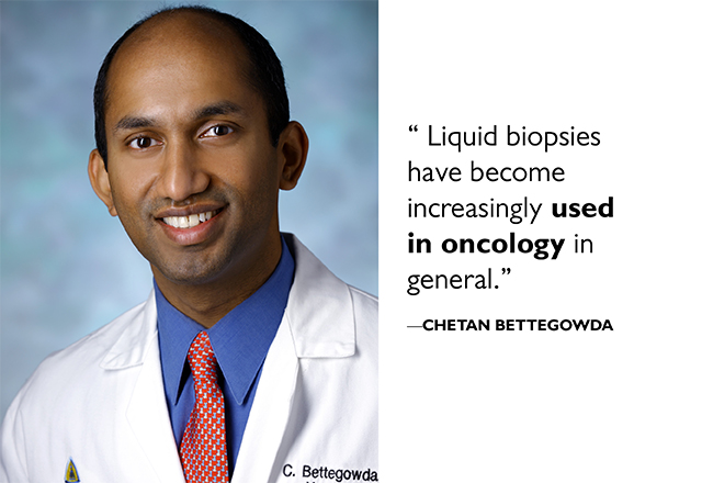
Liquid biopsies have become increasingly used in oncology in general, Bettegowda explains. Many cancer types have commercially available blood tests that track material derived from tumors. Johns Hopkins may be the leading institution in the world for liquid biopsy efforts, he adds, with several companies launched from technology developed here. However, no such tests have yet been approved for use in patients with brain cancer, leaving a critical gap in the field that Bettegowda and his colleagues are hoping to fill.
Before long, explains Bettegowda’s colleague Matthias Holdhoff, an oncologist at the Johns Hopkins Kimmel Cancer Center and collaborator on this work, the noninvasive liquid biopsy approach may allow the Johns Hopkins team to screen patients for the presence of brain cancers too minuscule to show up on imaging and then to customize treatments for each patient based on what’s most effective for a particular tumor type. In fact, Holdhoff and his colleagues are currently in the clinical trials stage of testing targeted therapies and experimental treatments that could be effective against brain tumors with specific mutations.
Moreover, some molecular features of tumors, captured through the liquid biopsy, will signal whether or not a patient might benefit best from surgery, chemotherapy, radiation or immunotherapy — or novel combinations of these therapies that researchers are currently developing at Johns Hopkins and elsewhere.
Being able to accurately characterize tumors could also allow doctors to better fine-tune treatment, Holdhoff says. “For select low-grade tumors, we could see an opportunity for initial close observation rather than pursuing early active treatment,” he adds. “We could give those patients more quality time, potentially for years, to be free of treatments that can be detrimental to quality of life.”
Avoiding Collateral Damage
Surgical and radiation oncologists at Johns Hopkins have also made great strides in avoiding collateral damage from treatment, Mukherjee says. In the last five years, he and his neurosurgical colleagues began implementing a device known as BrainPath, a tubular retractor that dramatically reduces the invasive nature of some surgeries to remove brain tumors.
Traditionally, the only way to reach a deep-seated tumor was through an open craniotomy, a surgery that involves large openings in the scalp, skull and dura — the membrane that covers the brain — and cutting through brain tissue to reach the cancer. The end result is often substantial blood loss, long hospital stays and recovery times, and an increased risk of damage to brain structures, which can cause neurological deficits.
Conversely, the new device (a tube with a diameter just over a centimeter wide) allows for a much smaller opening to access the brain. Rather than slicing through brain tissue, Hopkins neurosurgeons gently push the tissue aside using the tube, allowing them to resect entire tumors with minimal disruption to the surrounding brain structures, a gentler approach that helps preserve brain function.
The tubular retractor is currently only an option for patients with deep-seated, well-defined tumors and tumors that lie in and around the ventricles (the wide cavities inside the brain that produce cerebrospinal fluid). However, Mukherjee says, this option has significantly reduced the need for open craniotomies in brain cancer patients.
He and his colleagues now take a similar strategy when accessing tumors and removing them through the nose (what’s known as an endoscopic endonasal approach) by routinely using a device called SPIWay. Although the endonasal route spares patients from opening the skull, it comes with a significant risk of loss of taste and smell due to the trauma these procedures may cause to the nasal mucosa. During these multi-hour operations, instruments are constantly passed through the nasal passages, including elements to cauterize tissue, which often inadvertently burn away critical mucosa in the nose. SPIWay, a plastinated sheath that acts as guardrails to protect the nasal mucosa during surgeries, allows Mukherjee and otolaryngology–head and neck surgeon Nicholas Rowan to avoid these problems.
“A lot of these patients are young with decades of life left. If they can never taste red velvet cake again, or can’t smell smoke to save themselves from a house fire, it can be extremely detrimental to quality of life or even a safety issue,” says Mukherjee.
In other work, recently reported in the journal Neurosurgery, Mukherjee surveyed fellow neurosurgeons about their decision making between two different types of brain tumor surgery: supratotal resection, which removes the tumor along with a significant margin of adjacent tissue, versus gross resection, surgery that removes the tumor only.
The approach taken can have significant differences on quality of life, Mukherjee explains: While supratotal resection could give patients a longer time without tumor recurrence, it could also have a higher risk of harming neurological function. Gross resection preserves more healthy tissue, but could reduce quality of life in a different way if tumors recur and spread faster. Understanding how surgeons view which patients are eligible for each procedure provides a starting point for comparing and contrasting their outcomes, Mukherjee says.
The consensus of his crowdsourcing effort — that glioblastoma tumors in the right frontal and right temporal regions of the brain are candidates for supratotal resections — sets the stage for clinical trials to compare longer-term outcomes and quality of life that result from these two types of surgery.
First, Do No Harm
Sometimes understanding what it will take to improve quality of life for a particular patient with a brain tumor isn’t as simple as it seems, notes neuro-oncologist Karisa Schreck. One of her areas of research is studying dietary treatments that have shown some promise of eradicating cancer cells in the lab. Recently, she and her colleagues evaluated the feasibility of combining a ketogenic diet (high in fat, moderate in protein, and low in carbohydrates) and fasting in patients with high-grade gliomas. Both dietary treatments increase the production of ketones, a type of fuel made by the liver when cells are deprived of carbohydrates. In human cells in the lab and animal models, ketones have shown significant anticancer properties. The researchers wondered: Could eating foods that boost ketone production help slow, halt or even reverse cancer progression?
However, the diet patients needed to follow to even begin to answer this question was extremely restrictive, Schreck explains. “On non-fasting days, we had them count carbs and keep them under 20 grams per day. Even half a banana puts you over,” she says. On days when patients fasted, their only food was a 500-calorie ketogenic shake.
Although periodic blood tests and specialized MRIs showed elevated ketones in the bloodstream and brain, a factor that could have positive effects on the patients’ brain cancers, four of the 25 enrolled patients dropped out of the study early because they couldn’t comply or had side effects. Those who remained had trouble following the diet perfectly; it was a huge learning process to count carbohydrates and restrict their eating in such a specific way.
“The takeaway for me is that we think of things like dietary interventions as being relatively harmless, but they can have a very profound effect on quality of life. Patients might want more than anything to have a peach milkshake [while on the diet]. It’s hard for them to go to restaurants. They can’t have a glass of wine,” Schreck says. “These things are really important for some people and bring a lot of happiness, and taking them away can be harmful to quality of life, even if there’s a potential for extending survival.”
Schreck and other researchers now have their sights set on finding treatments that will mimic the effect of ketogenic diets without the negative impact on quality of life.
A Return to ‘Normal’
Banks finished chemotherapy in January 2021, and reports that she hasn’t needed any active therapy for her glioblastoma since then. Recently, she moved from having MRIs to monitor her brain cancer every two months to a three-month gap between scans — a reassuring sign that her disease remains stable with no new growth. Although she says she still gets “scanxiety” before each visit, her life remains much like it did before she was diagnosed, an important goal Banks had for her treatment.
“Everything is pretty normal. I do my work, I spend time with my family, I hang out with friends,” she says. “I’m grateful to have the quality of life I do today.”