Up to half of children with spina bifida whose defects are surgically repaired shortly after birth develop tethering, leading to later symptoms, including back and leg pain, weakness, and bowel and bladder dysfunction.
The traditional fix for these problems is an operation that loosens the spinal cord from the tissue that confines it. However, as a child grows taller and taller, this detethering procedure surgery must be performed repeatedly, causing a buildup of scar tissue that eventually causes its own set of issues.
Recently, pediatric neurosurgeon Mari Groves and her colleague Nicholas Theodore, director of Johns Hopkins’ Neurosurgical Spine Center, teamed up to offer patients a completely new approach: an operation that circumvents the tether completely and decreases tension on the spinal cord by shortening the spinal column instead.
Neurosurgeons have long performed procedures that inadvertently shorten the spinal column, such as partially or fully removing vertebrae when a cancerous tumor arises within the bone. However, to use this type of procedure for spinal cord tethering is new and extremely rare, says Theodore.
The team’s most recent patient exemplifies who would benefit most from the shortening procedure. By the time they performed the procedure in April 2019, the 20-year-old had already undergone six detethering operations; with each subsequent procedure, she benefited less and less. The pain and weakness wracking the patient’s legs and back severely limited the time she could spend on her feet. Bowel and bladder problems were steadily worsening.
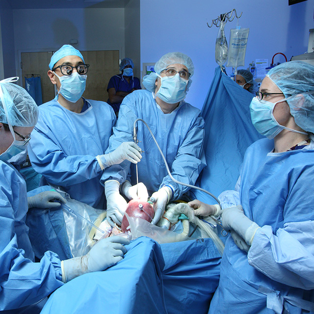
Rather than another detethering operation, she opted for the more durable solution. During the four-hour procedure, about 80 percent of the T12 vertebra was removed using image guidance and a robot that precisely places screws even in patients with very small pedicles. Under high-resolution ultrasound, Theodore, Groves and colleagues could see the spinal cord relax during the operation, transitioning from a tight stretch to a looser conformation that pulsated with each heartbeat. Theodore says this patient woke from surgery in tears of joy — although the pain of the procedure lingered, the excruciating pain from the tethered spinal cord had already dissipated.
The patient, says Theodore, is already running and hiking again, and recently began nursing school. “This is a significant departure in how we’ve traditionally treated these patients, but for the first time, it has the potential to be curative,” he says.