
How Do Neurons Talk to Each Other?
Shigeki Watanabe’s Lab Uses Electron Microscopy to Uncover How the Brain Sends, Processes and Stores Information
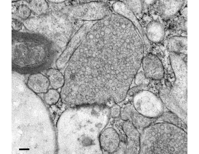
Human brain tissue collected during surgery and frozen 100 milliseconds after stimulation. Scale bar: 100 nm.
Chelsy Eddings
Neurons communicate in milliseconds, sending signals that allow the brain to process information, form memories and control behavior. Understanding how the tiny structures inside neurons make that possible means catching them in action.
Shigeki Watanabe, professor of cell biology at the Johns Hopkins University School of Medicine, uses electron microscopy to study these structures as neurons communicate and adapt. His team develops techniques that capture cellular activity at precisely defined moments, revealing processes that unfold rapidly in areas one-thousandth the width of a human hair.
One focus is how neurons replenish synaptic vesicles, the tiny bubbles that carry chemical messengers called neurotransmitters. Vesicles fuse with the cell membrane to release neurotransmitters into the synapse, the space between neurons. Neurons must then rapidly recycle vesicles to keep communicating. Genes associated with neurological disorders can affect this recycling and the energy metabolism that supports it, making Watanabe’s research important for understanding disease as well as healthy brain function.
Capturing the action
Synapses are too small to study in detail with light microscopy. Electron microscopy reveals their intricate architecture, but traditional methods require chemically fixing tissue, which preserves structure but makes it hard to capture what cells were doing at a particular moment.
An approach Watanabe’s lab helped develop, called zap-and-freeze, stimulates neurons and then freezes them at defined time points. The resulting images can be assembled into a kind of flipbook showing how structures change over time.
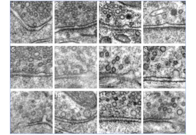
Using zap-and-freeze, Watanabe and his colleagues captured vesicles fusing with the cell membrane and traced how the membrane is retrieved to make new vesicles. The work helped resolve a decadeslong scientific debate. One model had proposed that neurons slowly build new vesicles using the protein clathrin, like sorting glass bottles by color by hand before melting them down to make new ones. Another suggested that vesicles simply pinch off from the membrane and are reused, like washing and refilling the same bottle.
“We found that neither model was correct,” says Watanabe. His team identified a new mechanism, called ultrafast endocytosis, that pulls the membrane back into the cell within milliseconds. The cell later forms new vesicles at endosomes, compartments that help sort and recycle cellular materials. It’s like sending bottles to a recycling facility for sorting, rather than taking the time to sort them by color at home.
Unexpected discoveries
“I have been fascinated by the many things hidden in electron micrographs,” Watanabe says. “A lot of research directions have started with looking through images and thinking, ‘I have never seen this before. What does it mean?’”
While studying motor neurons in C. elegans, a tiny worm, he noticed that axons, the long extensions that carry electrical signals between nerve cells, were not uniform tubes, as expected. They were beaded, with repeated swellings along their length. His team later saw the same pattern in mammalian neurons.
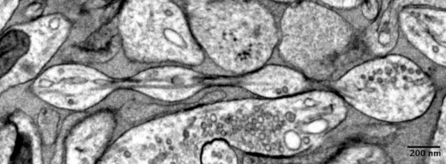
Axons wrapped in myelin, a fatty insulating layer, resemble strings of sausages, with small gaps between segments of myelin. But unmyelinated axons have traditionally been depicted as smooth tubes. Watanabe’s observations showed that unmyelinated axons can be beaded. This shape, known as pearling, may influence how electrical signals travel along these axons, and his team is exploring how.
The lab also studies microglia, immune cells Watanabe calls the brain’s “garbage collectors.” His team recently identified a mechanism by which microglia remove waste from neurons, helping maintain brain health.
The team is also applying its methods to human brain tissue left over from surgery, studying how disease-associated mutations affect vesicle recycling. Early comparisons of tissue from patients with epilepsy and Parkinson’s disease show differences they hope to explore further.
The next frontier
Watanabe often finds inspiration in publicly available electron microscopy data. By exploring these datasets, he can spot structures that raise new questions.
With unlimited resources, Watanabe says, he would build a system to generate large, 3D electron microscopy images of human brain tissue across diseases and genetic variations. Automated imaging and AI-assisted analysis would help produce detailed datasets to share with the global research community.
The goal would be to uncover structures and patterns scientists don’t yet know to look for, opening new avenues for understanding how the brain works and what goes wrong in disease.