
Following the Data
Legionella Protein Thought to Regulate Host Protein Synthesis Instead Targets a Metabolite

Photo by Kaile Gerard.
When Joshua Black joined the laboratory of Bloomberg Distinguished Professor Rachel Green at the Johns Hopkins University School of Medicine, he expected to study how a protein from the bacterium that causes Legionnaires’ disease disrupts protein production in infected cells. Instead, the data led him to a different cellular process: metabolism.
Black and colleagues found that the Legionella effector protein SidL chemically alters a molecule involved in glycolysis, the process cells use to break down simple sugars to make nutrients and energy. SidL had long been thought to interfere with translation, the cell’s process for making proteins, but Black’s early experiments suggested the effect was indirect.
As the project moved away from its original focus, it became a team effort involving 13 researchers across Johns Hopkins Medicine and beyond. They published the results in Molecular Cell in August 2026.
“Following the data and thinking creatively; those are both part of science,” says Black, a postdoctoral researcher. “It’s the art of science that people often overlook.”
Rethinking SidL
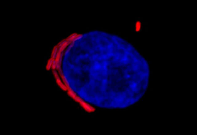
Legionella pneumophila naturally lives in water, where it grows inside tiny single-celled amoebae. When people inhale contaminated water droplets, the bacteria can reach the lungs and be taken up by immune cells called macrophages. Once inside, Legionella deploys an arsenal of roughly 300 effector proteins that interfere with normal cellular processes, helping the bacteria survive and multiply. Roughly three-quarters of these effectors are poorly understood.
Black initially studied SidL because of its presumed link to translation, an area of expertise for the Green lab. But early experiments showed that high levels of SidL turn off the mTORC1 cell-signaling pathway, which helps regulate translation in response to nutrients.
“That very quickly told me this is not really about translation,” Black recalls. “This is going to be several steps above it.”
Screening for a target
Black and the team set out to identify SidL’s substrate, the specific molecule it acts on. With help from A. Maxwell Burroughs in L. Aravind’s group at the National Institutes of Health (NIH), the team compared SidL to other proteins, which offered a clue: It most resembles LnaB, a different Legionella effector that transfers adenosine monophosphate, or AMP, onto host proteins.
But unlike LnaB, SidL did not appear to modify proteins, leaving its substrate elusive.
During a lab meeting, Katerina Romanov, a doctoral student in the bacterial pathogenesis lab of Johns Hopkins associate professor Tamara O’Connor, reminded Black that Legionella often requires unconventional thinking.
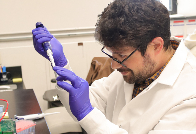
So Black looked beyond protein substrates to small molecules, testing SidL against a library of nearly 700 compounds that he acquired from Kevin Hicks’ lab at the University of Utah.
The screening identified SidL’s substrate as 3-phosphoglycerate, a small molecule produced during glycolysis. Follow-up experiments done with Edrees Rashan in Matthew Vander Heiden’s lab at MIT showed that SidL attaches AMP to it, creating a previously unknown compound called 2-AMP-3-phosphoglycerate. Subsequent experiments in mammalian cells showed that this disrupts glycolysis, which the researchers propose leads to the block in mTORC1 that in turn inhibits translation.
The team also found that SidL makes 2-AMP-3-phosphoglycerate in macrophages infected with Legionella, with the timing suggesting its synthesis primarily happens early during infection.
Building a team
As the project shifted toward metabolism, the researchers brought in additional expertise, including the Vander Heiden and Hicks labs. Some collaborations grew out of existing relationships; others began with a cold email.
“Having people recognize the lab and know it does good science means they’ll answer your email,” Black says.
At Johns Hopkins, Black worked closely with the O’Connor lab, which offered him the expertise and resources to perform Legionella infection experiments. When he needed help analyzing the chemical structure of 2-AMP-3-phosphoglycerate, he walked over to professor Caren Freel Meyers’ lab and found graduate student Charles Nosal eager to help.
“It was about managing a team, having rapport with people, and being able to say, ‘I have this problem. How can we solve it?’ and them responding, ‘This is how we solve it,’” Black says. “Without all of these people, this paper could not have happened.”
He says coordinating across labs and institutions was good preparation for one day leading a research group of his own.
“Learning how to spin up collaborations is a really useful skill,” Black says. “That’s part of what principal investigators do every day.”
Looking ahead
Black’s immediate next step is to continue collaborating with the O’Connor lab to understand how the newly discovered chemistry benefits Legionella. The findings also suggest SidL may be part of a broader family of bacterial effectors that use similar biochemical strategies. Ultimately, the researchers hope understanding this chemistry could inspire new tools for studying human health or biotechnology.
Black was fortunate to have both Green’s support and the funding stability to pursue a project that took an unexpected turn. That freedom has broadened how he thinks about approaching science, he says.
He intends to carry that approach into his next career move: identifying the right people, experiments and techniques to answer the questions that emerge, and then continuing to follow the data.
Other scientists who contributed to the work are Marco Catipovic and Catherine Valadez of the Johns Hopkins University School of Medicine. The project was supported by the Howard Hughes Medical Institute, NIH, the National Science Foundation and other sources.