
Making MRI Accessible: Sairam Geethanath’s Efforts in Low-Field MRI
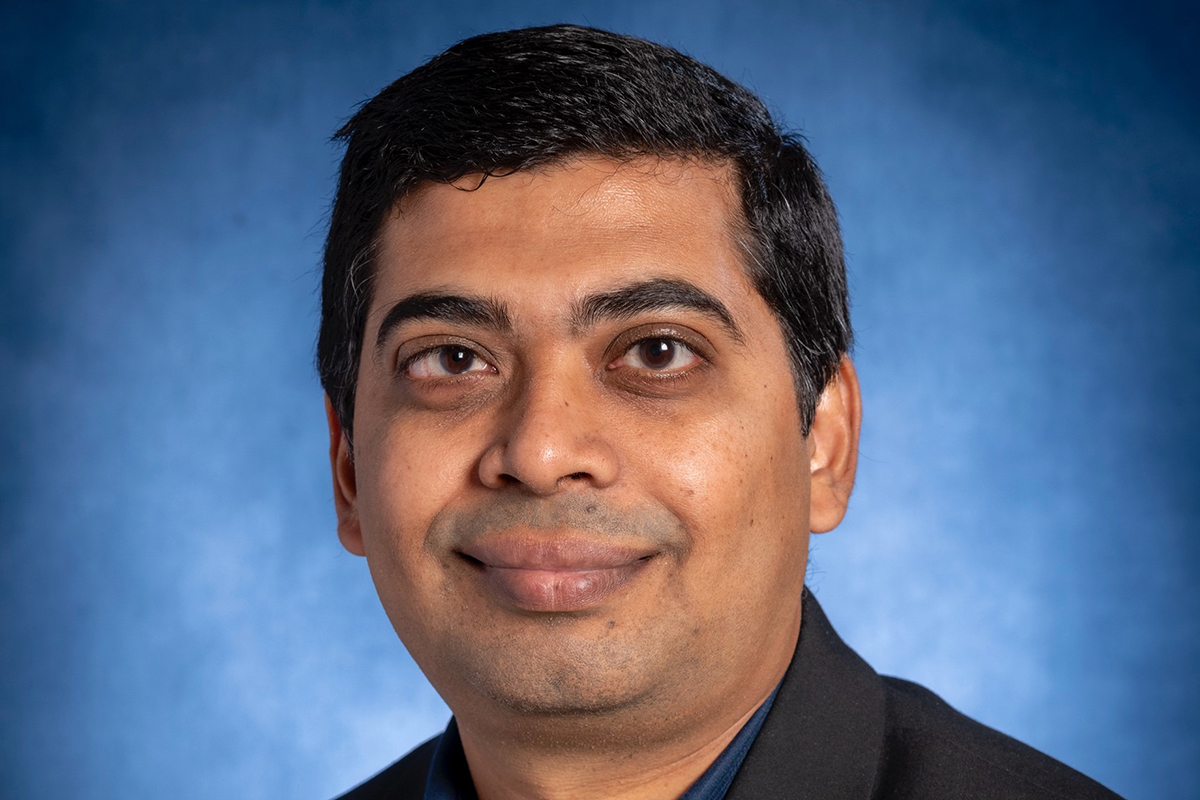
Since its introduction more than a half century ago, magnetic resonance imaging (MRI) has proven a lifesaving and safe tool. Multiple Nobel Prizes have been awarded for discoveries related to magnetic resonance, yet even today, two-thirds of the world has no access to this important modality. Sairam Geethanath, an associate professor of radiology, hopes to change that by developing and deploying low-field MRI scanners.
Geethanath, a biomedical engineer by training, became interested in low-field MRI while working in India in 2014. At that time, the Indian government, facing a ratio of less than one MRI scanner per million residents, pushed for the development of more scanners. As he worked on this, Geethanath became increasingly aware of the lack of MRI access across the globe. When he moved back to the United States, he realized the problem wasn’t limited to lower-income countries. Even in America, one of the countries with the highest proportion of MRI scanners per capita, rural areas still faced a huge lack of access to MRI.
Seeing an opportunity to help, Geethanath got to work. The field of MRI was full of researchers developing more powerful scanners — surpassing the traditional 1.5T (for Tesla, the unit for measuring MRI magnet strength) or 3T scanners to create 7+T research scanners. In MRI, more magnetic field strength means sharper images. These huge, expensive units produced highly precise images but were inaccessible to the billions of patients worldwide without MRI access.
What if he developed and adapted a smaller, cheaper, low-field MRI scanner (less than 1T in strength) that could produce clinically useful medical images?
Geethanath and his team were able to build functional low-field scanners as low as 0.05 T — or 60x weaker and much smaller and more portable — than a 3T scanner. However, the images it produced were too low-resolution to be used clinically. The answer, Geethenath realized, was in AI-assisted image reconstruction. He set to work developing AI tools to enhance the quality of the images. Today, using 0.05T MRI and AI image reconstruction, his lab can produce images of brain structures that match the resolution of 3T clinical images.
What makes Geethanath’s work truly unique is his lab’s comprehensive, end-to-end approach. Traditionally, MRI research has been siloed into labs focusing on either hardware development, image reconstruction or clinical applications. With his background as an engineer specializing in instrumentation and signal processing, however, Geethanath’s lab is one of the few in the world that is able to do everything in-house — from identifying a clinical need to building a low-field MRI scanner and developing the AI image reconstruction software needed to create clinically usable images.
Geethanath and team are deploying low-field MRI units to scan brain development, diseases and tumors, primarily in children ages 6 to 9.. They are focusing on this area due to their own expertise in brain MRI and because the units are currently too small to accommodate anything below the shoulders.
While clinical 1.5T and 3T scanners are large, taking up entire rooms and requiring structural floor reinforcement, low-field MRI scanners are much more portable, with the lowest-field scanners being small enough to fit on a tabletop.
Currently, Geethanath and his team are taking their technology from bench testing into the field. They have deployed low-field scanners to Fort Detrick to conduct scans for research. Additionally, healthy adult and pediatric volunteers have been scanned to refine the system before wider deployment.
Looking toward the future, Geethanath plans to take the technology into rural Maryland and surrounding regions. The goal is to provide these communities with low-cost MRI images to detect physical signs of serious medical issues such as brain disorders and diseases while they are still treatable.
“Once we are able to deploy these systems beyond the lab at Johns Hopkins and the local vicinity, we expect to have a profound impact on clinical interventions in the primary hospital setting globally,” Geethanath says.
By moving his work in low-field MRI work from the lab to the patient, Geethanath is working to ensure that, in the future, a patient’s location no longer determines access to imaging that could save their life.
For Clinicians Clinical Connection
Clinicians, discover the latest in research and clinical innovation from Johns Hopkins experts. Access educational videos, articles, CME courses and other resources from our world-renowned institution.